CASE NOTES
Acute Selenium Intoxication in Lambs
Case 2. Sudden Death, Neurological and Respiratory Signs in Merino Weaners Provided With a Water Supplement Containing Selenium
Elsa Glanville, Mackinnon Project, FVAS, University of Melbourne
Posted Flock & Herd August 2022
Introduction
Selenium is an essential trace element. Deficiency results in clinical disease and/or subclinical production loss following periods of profound dietary deficiency or a combination of low intake and high need (Hosking et al. 1986). It is typically seasonally and annually variable in presentation. The potential growth and production benefits associated with selenium have resulted in its inclusion in many routinely administered livestock products including drenches, vaccines, lick blocks and water supplements. However, excess selenium is not benign. Overdose can result in acute or chronic toxicosis. Acute toxicity in lambs results in cardiac failure causing lethargy, respiratory distress, neurological disturbance and death, mostly within 72 hours of exposure (Glenn et al. 1964; Blodgett & Bevill, 1987). Fatal dose rates of 1-2.2 mg/kg (oral: Lambourne & Mason, 1969; Caravaggi et al. 1970; Gabbedy, 1970) and 0.45-1 mg/kg (injected: Blodgett & Bevill, 1987) are reported. Given the regularity with which selenium is included in sheep products, several overdose scenarios are possible. The case below illustrates the consequence of providing surplus selenium via an oral supplement.
History
In mid-December 2021 near Bathurst NSW, two mobs of September-drop Merino lambs were weaned. The lambs were 3-4 months old. They were weighed and drafted into a 'light' mob <18 kg (n = 470) and a 'heavy' mob >18 kg (n = 630). All were vaccinated (5-in-1 only), drenched (Startect®, to max weight in mob) before being placed in containment yards for weaning. They were provided withad lib. lucerne hay and were started on 50 g/head/day pellet/barley mix. The pens contained a mixed sward of grasses and weeds. Water was provided from a bore to high-flow, low-volume troughs, one in the light pen and two in the heavy pen, to which 10 ml/head/day liquid mineral supplement containing 1500 mg/L sodium selenate was added (intended to be added for three days after weaning).
The producer noted intermittent lamb deaths pre-weaning, including several lambs found after imprint feeding. Twenty-four hours after weaning, four lambs were found dead across both pens, with no obvious external abnormalities. The next day, the producer noticed increasing numbers of lambs frothing from the mouth and nares, panting, walking with a stilted or 'proppy' gait and another dead with froth around the nares. Seven more dead lambs were found in the 'light' mob whilst one more was found in the 'heavy' mob. A private veterinarian was called to investigate, suspecting a toxicity.
EXAMINATION OF LAMBS IN WEANING YARD
At least six lambs with respiratory distress, lethargy and/or neurological signs were noted in each mob. These lambs were tachypneic, with froth at the mouth, pink mucous membranes with rapid refill (Figure 1). They typically stood with a high head carriage and walked with a stiff, stilted gait. Unaffected lambs appeared calm, bright and alert.


Figure 1. Typical presentation of live clinical cases included lambs in various degrees of respiratory distress with a stable foam around the mouth (and nares, not shown in this case).
Necropsy findings
A clinical examination was performed on a moribund lamb from the 'light' mob that was subsequently necropsied. Five recently deceased lambs were also presented for necropsy. In total, three lambs were presented from each mob. Unfortunately, samples were unable to be taken for histopathology at the time of the necropsies. However, fresh samples were taken and confirmed the diagnosis.
'Light' mob lambs
Lamb 1: 10 kg emaciated, non-responsive lamb. Normal respiratory excursions. Humanely euthanased by producer. Necropsy revealed an extensive abscess extending from Gudair® vaccine site on right neck proximal to base of ear. Purulent material tracked through fascial planes into atlas/axis, extending cranially and caudally through spinal canal. The carcass was malnourished, but the gross necropsy was otherwise within normal limits.
Lamb 2: small lamb dead in lateral recumbency (Figure 2). White foam from both nares. No other external abnormalities. Carcase in fair condition. Diffuse muscle pallor with flabby texture, most notable in hindlimbs. Abundant straw-coloured fluid in pericardial and pleural cavities. Stable foam through trachea to the bifurcation. Lungs heavy and wet, lesions consistent with pulmonary oedema. Cardiac muscle was mottled with areas of pallor.
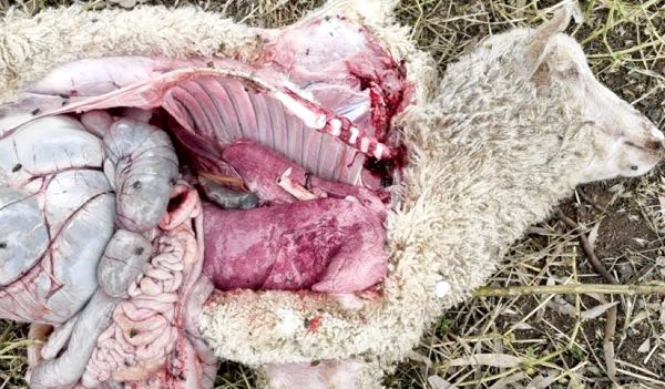

Figure 2. Light draft lamb found dead with froth at nares. Display stage of necropsy is presented, showing congested, wet lungs with evidence of pulmonary oedema. There was diffuse skeletal muscle pallor and discolouration of the heart muscle. Death occurred several hours prior to necropsy on a hot day.
Lamb 3: light lamb in poor body condition. No obvious external abnormalities. Gelatinous atrophy of pericardial and perirenal fat, blunt rumen papillae and little rumen fill. No other gross abnormalities. A most likely diagnosis of malnutrition was made, but fresh liver and kidney samples were taken.
'Heavy' mob lambs
Lambs 4 and 5: heavy lambs, good condition, dead in lateral recumbency. Similar to lamb 2 with foam from nares and no other external abnormalities. Abundant straw-coloured pleural, pericardial and peritoneal fluid. The main bronchi and trachea were filled with stable foam. Lungs were congested, heavy and wet, with lesions consistent with pulmonary oedema. Skeletal muscles were pale with a flabby texture, especially through the hindlimbs. The liver of each lamb was congested and moderately enlarged (Figure 3). The heart of lamb 4 was noticeably discoloured, with regions of pallor extending out from coronary arteries (Figure 4). Fresh liver and kidney samples were taken.


Figure 3. Lambs 4 (A) and 5 (B), heavy draft lambs found dead with froth from nares. Images from the display stage of the necropsy show signs consistent with lamb 2; wet lungs and evidence of pulmonary oedema including interlobular oedema, straw-coloured pleural fluid and diffuse muscle pallor. Livers were congested with moderate hepatomegaly.
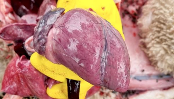
Figure 4. Heart of lamb 4 showing areas of pallor extending from the coronary vessels.
Lamb 6: dead more than four hours at necropsy. Carcass in good condition. Abdominal distension. Serosanguinous peritoneal and pleural fluid. Ecchymotic haemorrhages over ventral serosal surface of rumen and abomasum, with discoloured fluid-filled loops of bowel (Figure 5). Rumen distended with foetid fluid and barley, mucosa sloughing to reveal haemorrhagic areas (care interpreting sloughing mucosa given time since death; pH not tested due to time since death). Abomasal mucosa inflamed, contents included whole barley. Based on gross necropsy, a provisional diagnosis of ruminal acidosis was made. Liver and kidney were sampled.

Figure 5. Heavy draft lamb (lamb 6) died several hours prior to necropsy. Image from display stage of necropsy, showing gross lesions inconsistent with lambs 2, 4 and 5. This lamb is suspected to have died from ruminal acidosis, rather than intoxication.
The preliminary diagnosis from the gross necropsy findings and clinical signs was selenium intoxication. The time that had elapsed between initial access to the likely source of the excess selenium and the presentation of signs, 12-24 hours, was consistent with previous investigations of oral selenium intoxication (Tiwary et al. 2006).
Laboratory Findings
Fresh liver samples were submitted for selenium and copper tissue concentrations based on a strong suspicion of selenium intoxication and a potential for high levels of copper intake based on amount of supplement potentially ingested. Although kidney samples were taken, they were not analysed as the results from the liver were diagnostic.
| SAMPLE | LIVER COPPER | LIVER SELENIUM |
|---|---|---|
| REFERENCE | 0.23-3.67 mmol/kg wet wt | 1.3-19 umol/kg wet wt |
| LAMB 2 | 0.89 | 164 H |
| LAMB 3 | 0.65 | 4.8 |
| LAMB 4 | 1.5 | 153 H |
| LAMB 5 | 0.47 | 102 H |
The laboratory findings confirmed that the cause of death of the lambs that presented with a history of sudden death, respiratory and neurological signs was selenium intoxication. Based on the management history, the intoxication occurred due to excess intake of supplement provided in the water troughs. The likely amount of selenium ingested relative to toxic levels is discussed below.
The cervical abscess, ruminal acidosis and malnutrition cases highlight the importance of conducting a suite of necropsies when presented with a mortality outbreak, especially in weaners when multiple disease processes may be occurring contemporaneously in the mob.
Discussion
Here we report acute, fatal selenium intoxication causing cardiac insufficiency, resulting in tachypnea, respiratory distress, skeletal muscle damage and/or sudden death (Glenn et al. 1964, Tiwary et al. 2006). This case of intoxication from excess mineral supplement added to the water source demonstrates the ease with which young sheep can be accidentally overdosed with fatal consequences.
The liquid supplement had been previously used by this operator with no complications. However, the producer had started using an in-house weaning protocol with a typographical error. The dose of the liquid supplement exceeded the label recommendation (label: lambs <30 kg, 5 ml/head over 3 days; protocol: 10 ml/head for 3 days). The label instructions suggested the total volume each day be added to troughs either once or twice a day. Having used the product previously, the producer elected to add it once daily. Hence, at a rate of 10 ml/head/day, 4.7 L (470 head) was added to the trough in the light draft pen on day one of weaning. A total 7050 mg of Se (1500 mg/L Se in supplement) was added to the 40 L trough, resulting in an initial concentration of 176 mg/L. As water was drawn out of the trough, that concentration would have decreased. However, as Se intoxication in lambs has occurred with oral doses of 1-2.2 mg/kg, or 18-40 mg for an 18 kg lamb, the initial concentration easily exceeded that dose. On a warm day, a lamb may drink 5-10% bodyweight, 0.9 L for 18 kg lamb. On this occasion, 0.9 L would have contained 158 mg selenium, or 8.8 mg/kg for that 18 kg lamb. Hence, early drinkers may have received over four times the toxic dose. In the heavy draft, the total dose of the liquid supplement was divided between two troughs, with 4725 mg selenium added to each and an end concentration of 118 mg/L. At a similar level of water ingestion, the early drinkers in the heavy mob would have ingested over two times the toxic dose. The lower selenium concentration in the water and the heavier lambs may explain the lower mortality in the heavy mob. Selenium administered via the oral route may be more available to young lambs, likely over-represented in the light mob, due to incomplete rumen development.
During the initial phone call discussing the case, the producer was advised to remove access to the supplement in case of any related toxicity. Hence the troughs were immediately dumped and flushed with clean water. It was also recommended that subsequent doses of the supplement be avoided. It is likely that these recommendations reduced the number of mortalities. We also recommended that the lambs be monitored for further cases as historical case reports suggest most cases develop over the first 72 hours but some present five days or more after last access or administration. No further cases were observed four days after the initial presentation.
In addition to the lambs that died peracutely, there were lambs with less immediately fatal respiratory and neurological signs. No specific antidote to selenium intoxication is available, so the recommendation was made to manage symptoms and avoid stress, excess exercise and provide fresh water and good roughage. Despite this, six lambs with clinical signs died after several days (three from both mobs). A total of 20 lambs died with signs consistent with selenium intoxication (1.8%). Due to other concurrent disease processes, this mortality rate is a best estimate. The producer was unsure of the total showing signs but felt that those with less severe respiratory signs recovered well. There are reports of sublethal selenium intoxication resulting in scouring and pneumonia after pulmonary damage. No such signs were seen in this case.
With the consent of the producer, the manufacturers of the liquid supplement were notified of the details of this case. The instructions for use of the supplement in lambs have subsequently been modified and now include a discussion of the risks of adding the supplement to low-volume, high-flow troughs and a recommendation to further split doses to reduce the risk. This case highlights the importance of considering the dilution effect when providing supplements to livestock via water sources. As an additional note, fortunately these lambs were not also provided with an additional source of selenium, via either the drench or vaccine used at weaning, as is often the case. More deaths may have occurred if they had been.
This highlights the need for producer awareness of the risk of selenium intoxication, especially in young sheep at times of increased risk. The risk of toxicity increases during times of stress, including lamb marking and weaning, in young sheep with incompletely developed rumens and in those with concomitant disease, where selenium transport and storage may be reduced. For veterinarians, this case serves as a reminder to include selenium intoxication as a differential for respiratory distress, lethargy, neurological signs and sudden death especially where a stable foam is noted in the trachea and around the mouth and nares. Key diagnostic samples include fresh liver for selenium concentration and liver, lung and heart muscle for histopathology. Preventing access to the supplement and ensuring no further supplementation occurs over the following month (for short-acting sources) will prevent subsequent mortalities in survivors.
References
- Blodgett DJ, Bevill RF (1987). Acute selenium toxicosis in sheep. Veterinary and Human Toxicology 1987;29:233-236
- Caravaggi C et al. (1970). Experimental acute toxicity of orally administered sodium selenite in lambs. Research in Veterinary Science 11:146-149
- Gabbedy BJ (1970). Toxicity in sheep associated with the prophylactic use of selenium. Australian Veterinary Journal 46:223-226
- Glenn MW, Jensen R, Griner LA. Sodium selenate toxicosis: pathology and pathogenesis of sodium selenate toxicosis in sheep. American Journal of Veterinary Research 1964;25:1486-1494
- Hosking WJ et al. (1986) Selenium. In: Hosking WJ et al. editors. Trace Elements for Pastures and Animals in Victoria. Victorian Government Printing Office, Department of Agriculture and Rural Affairs, Melbourne, Victoria, Australia, 20-26
- Lambourne DA, Mason RW (1969). Mortality in lambs following overdosing with sodium selenite. Australian Veterinary Journal 45:208
- Tiwary AK et al. (2006) Comparative toxicosis of sodium selenite and selenomethionine in lambs. Journal of veterinary diagnostic investigation 18:61-70.