CASE NOTES
Degenerative joint disease of the stifle joints in mature Angus cows
Bruce Watt, Central Tablelands Local Land Services, Bathurst and Anthony Chamings, Elizabeth Macarthur Agricultural Institute, Menangle
Posted Flock and Herd August 2025
INTRODUCTION
Degenerative joint disease (DJD), usually affecting the hip, stifle and hock, is quite common in older beef cows (Parkinson et al., 2019). In the experience of the first author, the stifle joints are most commonly affected. DJD is a significant cause of wastage in mature cows on some properties on the Central Tablelands. Most affected cows become severely lame on one or both hindlimbs, lose weight and are either sold as culls or destroyed on farm as they become emaciated and unfit to load.
In this case report, a Central Tablelands cattle producer reported an unusually high number of cases of DJD in his cows.
HISTORY
Over the previous 4-5 years, a producer running approximately 600 Angus cows had seen approximately 15 mature Angus cows that had developed what was presumed to be DJD of the stifle joints. At the time of the investigation, the owner had five cases, mostly in 7-to-9-year-old cows. One cow became so thin and immobile that she was euthanased and subsequently necropsied. Both stifle joints were submitted to EMAI.
CLINICAL FINDINGS
The affected cow, a mature 7-9-year-old Angus cow, was bright and alert but emaciated and reluctant to walk (Figure 1). When pushed the cow walked slowly and gingerly with reluctance to bear weight on the left hind leg (Video 1). She had a markedly enlarged left stifle and moderate enlargement of the right stifle.
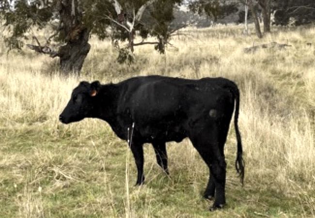
Figure 1. Case 1, a barely mobile mature Angus cow with enlarged, crepitant stifle joints
Video 1. Gait of affected cow
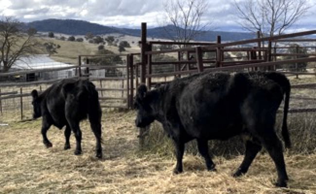
Figure 2. Two cows with degenerative joint disease of the stifle joints
NECROPSY FINDINGS
The cow was in thin body condition. Both stifles were grossly enlarged with excess synovial fluid in distended joint capsules. Both stifles had significant peri-articular exostoses and erosions of the articular surfaces (see additional details below).
RADIOLOGY FINDINGS
The right stifle was removed and radiographed at a local private veterinary clinic (Figure 3). There were moderate amounts of bony proliferation along the margins of the femur, tibia and patella in the stifle and there were several small, irregularly shaped pieces of bone in the surrounding soft tissue.
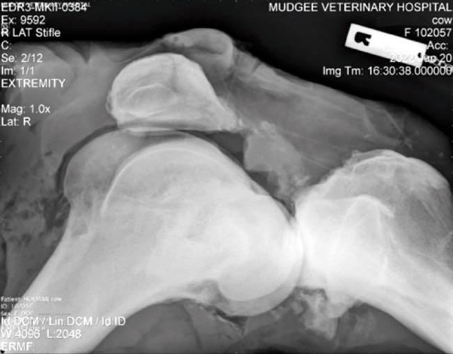
Figure 3. Radiograph of the right stifle of case 1
LABORATORY FINDINGS
A sample of the joint aspirate was submitted to the University of Sydney Diagnostic laboratory via the Elizabeth Macarthur Agricultural Institute (M22-09274). The aspirate was a pink-yellow colour and moderately cloudy. It contained 30 g/L of protein and there was a mildly elevated total nucleated cell count consistent with a mild inflammatory joint effusion. The sample was PCR negative for both Chlamydia pecorum and Mycoplasma spp.
Both stifles were submitted for post-mortem investigation. In the left stifle, on the medial condyle of the femur there was an irregular, approximately 2cm x 3cm, full thickness loss of the articular cartilage, and the exposed subchondral bone was haemorrhagic (Figure 4). The haemorrhage extended 8-10mm into the underlying bone (Figure 5). There was roughening of the cartilage on the medial and lateral trochlear ridges and on the articular surface of the patella. The cartilage over the surface of the medial tibial plateau was moderately roughened with several areas of yellow-brown discolouration (interpreted as cartilage thinning) (Figure 6).
In the right stifle, over the medial condyle of the right femur, there was erosion (almost full thickness) of most of the cartilage (Figure 7) and a 1cm round area of full thickness loss of the cartilage at the cranial margin of the condyle. There was severe roughening of a 2cm x 1cm area of cartilage of the midpoint of the lateral trochlear ridge and moderate roughening extending along the lateral ridge and into the trochlear groove of the femur (Figure 8). There was multifocal roughening of the articular surface of the patella. There was a large irregular area of significant erosion/ulceration of the articular cartilage over the right medial tibial plateau (Figure 9).
The synovium around the margins of the joints was reddened and oedematous and elsewhere it was irregularly thickened (Figure 4 and Figure 7). There were numerous chondrophytes/osteophytes fused to the margins of the articular cartilage (Figure 10). Occasional chondrophytes/osteophytes were also found in the synovium (Figure 6). Cruciate, patella, meniscal and collateral ligaments in both joints grossly appeared intact. There was some fraying of the cranial cruciate of the left stifle, however it was unclear whether this was artefactual from when the joint was reflected open. Based on the gross findings a diagnosis of degenerative joint disease was made in both stifles. The disease was graded as the most severe category of "chronic" based on the grading system in Zachary and McGavin (Olson and Carlson, 2017).
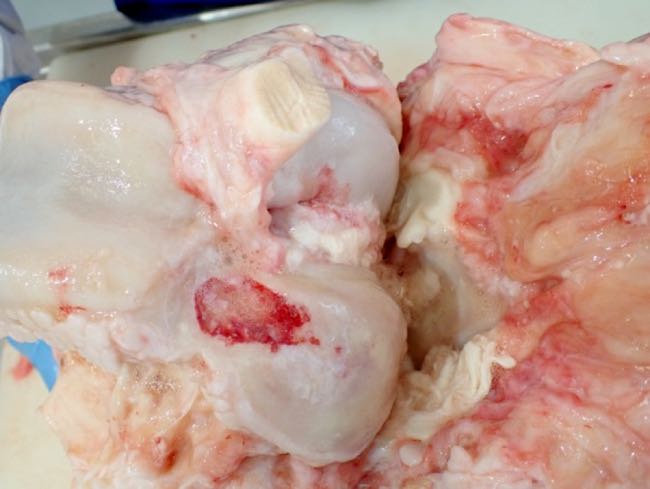
Figure 4. The articular surface of the left femur showing a large full-thickness ulceration of the cartilage over the cranial medial condyle surrounded by roughened cartilage. Roughened and reddened synovial tissue is visible in the left periphery of this image.
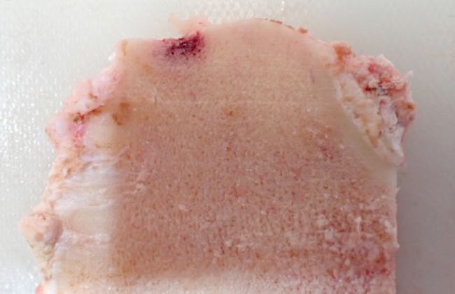
Figure 5. Cross section of the medial condyle of the left femur, showing haemorrhage extending into the subchondral
bone beneath the erosion shown in Figure 4
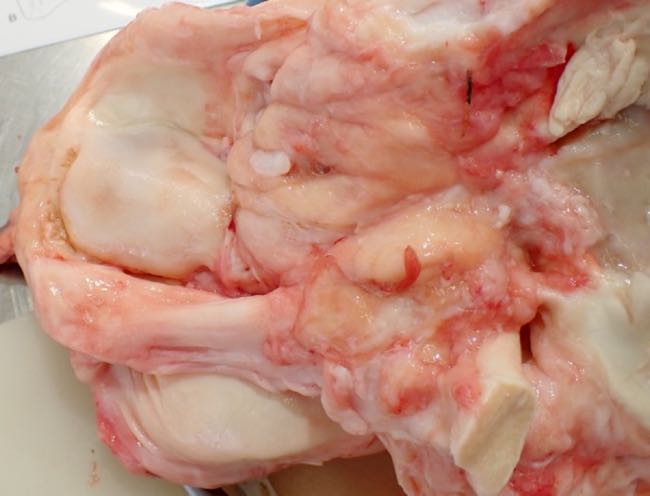
Figure 6. Left tibia showing thinning and roughing of the cartilage of the medial tibial plateau. Occasional
osteophytes are visible in the synovium (arrow heads).
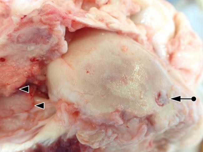
Figure 7. Erosion and roughening of the cartilage over the medial condyle of the right femur. A 1cm area of
ulceration is visible at the cranial margin of the medial condyle (arrow). Variable thickening of the synovium is
also visible (arrow heads).
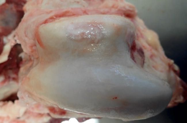
Figure 8. Roughening of the cartilage of the lateral trochlear ridge and trochlear groove of the right femur
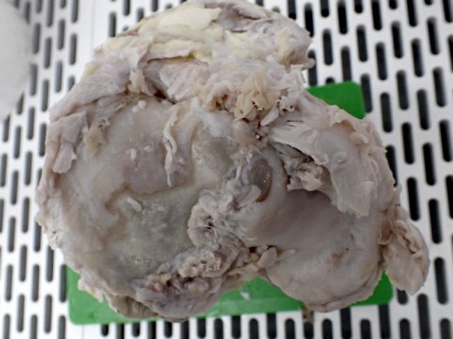
Figure 9. Severe erosion/ulceration of the articular cartilage of the medial tibial plateau of the right tibia
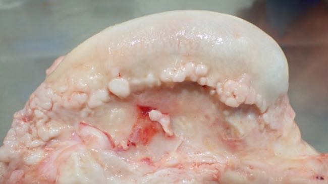
Figure 10. Numerous chondrophytes/osteophytes are fused along the margin of the articular surface of the medial
femoral trochlear ridge
DISCUSSION
The changes identified in this animal illustrate the types of gross changes present in advanced DJD in cattle. The DJD was clearly interfering with this animal's ability to move around and feed, ultimately resulting in the cow losing body condition. Several authors have identified DJD as causing economic losses for producers through poor reproductive performance in affected bulls (Persson, Söderquist and Ekman, 2007), loss of body condition score and reduced production of meat and milk (Barbosa, Lima and Belo-Reis, 2014). More broadly, DJD would contribute to the overall amount of lameness seen in beef herds. Lameness was the third-most frequent class of disease reported in a survey of American beef cattle herds (USDA, 2020), amongst the top three diseases for veterinary expenditure in beef cattle in the US (Hird et al., 1991) and the seventh-most common reason for mortality or culling of beef cattle in Western Australia (Aleri et al., 2021).
DJD is a relatively common condition affecting many species, including humans, dogs, horses and cattle. It is a progressive disease characterised by the destruction of articular cartilage leading to alterations of the subchondral bone and periarticular soft tissue and ligaments (Coleman et al., 2020). Sometimes there is a clear predisposing cause (e.g. trauma, joint infection, conformational deformity) resulting in the development of osteoarthritis (secondary osteoarthritis/DJD) but in many cases the initiating cause is unknown (primary osteoarthritis/DJD). Two major factors are thought to be relevant to the development of osteoarthritis - mechanical load and rate of tissue repair/catabolism. Excessive load, e.g. due to an abnormal conformation or excessive use, could overwhelm a normal joint's ability to repair itself. Similarly a joint under normal load experiencing increased catabolic stimuli (e.g. increased proteolytic enzyme production e.g. matrix metaloproteases, by chondrocytes, synoviocytes or neutrophils in response to inflammation) may also result in loss of articular cartilage (Coleman et al., 2020). Severe trauma such as a fracture or ligament rupture or tearing was not seen in either joint in this cow, but less catastrophic trauma earlier in life could not be entirely excluded. Trauma was the most common injury seen in the stifle of cattle in one retrospective study (Newcomer and Chamorro, 2016).
Initial stages of DJD affect the articular cartilage and can be appreciated grossly as the cartilage taking on a dull, yellow, granular appearance, which may feel soft when palpated (Shupe, 1961; Coleman et al., 2020). This degeneration progresses to erosion of the cartilage where small clefts or fissures may appear. The cartilage then takes on a frayed or split appearance before the most advanced stages of the disease in which the cartilage is completely eroded and the underlying subchondral bone is exposed. Over time the bone will become smooth (eburnation) or grooved, the surrounding joint capsule thickened and osteophytes will develop (Shupe, 1961; Olson and Carlson, 2017). The stifle joints of this animal had many features of this latter chronic stage of DJD but had not yet developed eburnation of the underlying bone.
In cattle, several predisposing risk factors have been proposed as contributing to the development of primary DJD. These risk factors include breed/hereditary factors (Shupe, 1961; Dutra, Carlsten and Ekman, 1999), hindlimb conformation (Shupe, 1961), osteochondrosis or growth plate changes when young (Dutra, Carlsten and Ekman, 1999; Heinola et al., 2006) and mineral deficiencies (Heinola et al., 2006). A survey of young 12-month-old beef bulls in Sweden found that hindlimb joint pathology such as osteochondrosis in the stifle is common (Dutra, Carlsten and Ekman, 1999) and this condition has been proposed to potentially develop into DJD later in life. In the case described in this paper, there were no obvious osteochondrosis lesions in either stifle.
In Angus cattle, several recent reports suggest that the incidence of lameness in grain-fed Angus cattle has increased over the last 20 years (Grandin, 2024). Some degree of lameness was observed in 8.2% of Angus cattle assessed in slaughter yards in the United States (Davis et al., 2024), which some authors have attributed to a combination of hoof conformation in addition to the increase in animal weight and size relative to 20 years ago (Grandin, 2024). With the importance of soundness to cow performance, Angus breeding associations have in place foot and leg conformation scores to help producers determine which animals should enter breeding programs and which should be culled (Angus Australia, 2025). The need to further investigate the hoof, foot and leg and their impact on the productive life of Angus cows and bulls has been identified as a research priority by some authors (Giess et al., 2021). Such research might better characterise the incidence of types of lameness, including DJD, in this breed.
The underlying cause of the arthritis seen in this individual and others on the one property was not determined. No infectious agents were found on testing of the joint fluid. Foot and hindlimb confirmation did not appear to play a role in the disease seen in this cow. This case and the other similar cases on this property were presumed to be primary DJD as they were bilateral with no evidence of ligament damage or fractures.
Anecdotally, DJD is a low-prevalence disease in mature Angus cows and bulls on the Central Tablelands, though is rarely observed in other breeds and in cattle on the western slopes and plains. This observation requires substantiation.
ACKNOWLEDGEMENTS
I would like to thank Jack Holman, then at the Mudgee Veterinary Hospital for taking the radiographs.
REFERENCES
- Aleri JW et al. (2021) A descriptive retrospective study on mortality and involuntary culling in beef and dairy cattle production systems of Western Australia (1981-2018) Australian Veterinary Journal 99(9):395-401 doi.org
- Angus Australia (2025) Collecting Structural Soundness Scores www.angusaustralia.com.au (Accessed: 6 May 2025)
- Barbosa JD, Lima DHS and Belo-Reis AS (2014) Degenerative joint disease in cattle and buffaloes in the Amazon region: a retrospective study Pesquisa Veterinária Brasileira 34 doi.org
- Coleman MC et al (2020) Chapter 38 - Diseases of the Bones, Joints and Connective Tissues, in BP Smith, DC Van Metre, and N Pusterla (eds.) Large Animal Internal Medicine (Sixth Edition) St. Louis (MO): Mosby, pp. 1197-1266.e14 doi.org
- Davis MK et al. (2024) Benchmarking current preslaughter management factors, welfare indicators and meat quality outcomes at commercial fed cattle processing facilities in the United States Translational Animal Science 8 p. txad150 doi.org
- Dutra F, Carlsten J and Ekman S (1999) Hind limb skeletal lesions in 12-month-old bulls of beef breeds Zentralblatt Fur Veterinarmedizin. Reihe A 46(8):489-508 doi.org
- Giess LK et al. (2021) Genetic parameter estimates for feet and leg traits in Red Angus cattle Journal of Animal Science 99(11) p. skab256 doi.org
- Grandin T (2024) Problems with Congestive Heart Failure and Lameness That Have Increased in Grain-Fed Steers and Heifers Animals: an open access journal from MDPI 14(19):2824 doi.org
- Heinola T et al. (2006) Consequences of hazardous dietary calcium deficiency for fattening bulls Acta Veterinaria Scandinavica 48(1):25 doi.org
- Hird DW et al. (1991) Expenditures for veterinary services and other costs of disease and disease prevention in 57 California beef herds in the National Animal Health Monitoring System (1988-1989) Journal of the American Veterinary Medical Association 198(4):554-558
- Newcomer BW and Chamorro MF (2016) Distribution of lameness lesions in beef cattle: A retrospective analysis of 745 cases The Canadian Veterinary Journal = La Revue Veterinaire Canadienne 57(4):401-406
- Olson EJ and Carlson CS (2017) Chapter 16 - Bones, Joints, Tendons and Ligaments. in JF Zachary (ed.) Pathologic Basis of Veterinary Disease (Sixth Edition) Mosby, pp. 954-1008.e2 doi.org
- Parkinson TJ et al. (2019) Diseases of cattle in Australasia. A comprehensive textbook 2nd Edition. Aukland: Massey University Press. pp. 974-5
- Persson Y, Söderquist L and Ekman S (2007) Joint disorder; a contributory cause to reproductive failure in beef bulls? Acta Veterinaria Scandinavica 49(1):31 doi.org
- Shupe JL (1961) Arthritis in Cattle The Canadian Veterinary Journal = La Revue Veterinaire Canadienne 2(10):369-376
- USDA (2020) Beef 2017, Beef Cow-calf Health and Management Practices in the United States, 2017, report 2 Fort Collins, CO: USDA-APHIS-VS-CEAH-NAHMS www.aphis.usda.gov