CASE NOTES
Hemlock Poisoning of Wagyu steers in the Northern Tablelands
Dr Samantha Boland BVSc, District Veterinarian, Inverell Local Land Services, Dr Meg Parsons BVBiol BVSc, District Veterinarian, Glen Innes Local Land Services and Dr Andrew Biddle, District Veterinarian, Inverell Local Land Services
Posted Flock and Herd July 2023
Summary
Poison hemlock (Conium maculatum) is a biennial herbaceous plant that is highly toxic to livestock and humans. Reported cases of poisoning in livestock within the literature are uncommon, which may be due to the tendency of animals to avoid grazing the plant because of unpalatability (McKenzie, 2012). Stock may graze areas containing hemlock without incident, signifying that there may be additional factors such as lack of alternative forage that would drive them to consume the plant. Cases may also be underreported due to misidentification, as the plant closely resembles other less toxic weeds such as Queen Anne's lace (Daucus carota) and bishop's weed (Aegopodium podagraria).
History
In late January 2023, west of Inverell, NSW, approximately 200 two-year-old Wagyu steers were rotated into a new paddock of primarily lucerne, with the remainder (approximately 15%) comprised of assorted native- and paspalum-dominant pasture. There had been no recent chemical application on this paddock or those adjacent. Weed species identified by the stock owner included St Barnaby's thistle (Centaurea solstitialis), Queen Anne's lace (Daucus carota), purple top (Verbena spp.) and billy buttons (Craspedia spp.). Water supply was a single trough from a reliable bore that also supplied other paddocks and residential water. They were supplemented with a commercial mineral lick block, and the trough treated with commercial bloat oil. The steers were introduced onto the property in winter 2022 and had received a clostridial vaccination prior to their arrival.
The cattle, pasture and water supply were monitored daily by a dedicated stockperson. The pasture was also assessed by an agronomist who determined that, while pasture mass was significantly decreased, the paddock could be grazed for an additional three days prior to the next rotation.
On day five of the paddock rotation, a staff member observed from a distance that stock appeared to be unsettled and fence walking. Upon further investigation by the primary stockperson, approximately one quarter of the stock were found to be extremely agitated and running through fences, with deceased animals on the fence line. Upon checking the sole trough in the paddock, approximately nine animals were found either recumbent and moribund, or dead.
Clinical Signs and Case Progression
Additional clinical signs included hypersalivation, ataxia, muscle fasciculations, dyspnoea, tachypnoea, regurgitation of rumen fluid, and seizures. Mentation varied from disorientated, to lethargic, to comatose. Over the next 24 hours, eight of the severely affected steers died or were euthanised. One animal had blood collected (Steer 1) and three animals were necropsied by feedlot staff (Steers 2, 3 and 4). Animals that were recumbent but could be roused were examined by numerous stockpersons, with elevated rectal temperature a common finding. The feedlot's consultant veterinarian was notified who advised preliminary treatment with oxytetracycline, dexamethasone and vitamin B complex.
An additional 30 head of cattle were less affected and recovered rapidly. However, they continued to display lethargy and shade seeking behaviours. They were moved to a new paddock and subsequently improved over the next week.
Three days after the initial insult, Local Land Services staff were engaged to assist with the disease investigation. On physical examination, two steers (Steers 5 and 6) displayed sternal recumbency, tachypnoea, dyspnoea, dehydration, reduced gut sounds, decreased menace response, dull-to-obtunded mentation and muscle fasciculations. These animals had blood collected at this time. Another deceased animal (Steer 7) was necropsied and then Steer 6 was euthanised and necropsied. A paddock walk was also undertaken at this time to ascertain if any potentially toxic plants were present.
Eleven days after the onset of clinical signs, further blood sampling was undertaken on steers from four separate groups to investigate if a pre-existing health condition had led to the incident and to assess if treated animals had ongoing health effects. These groups included members from the affected mob that had received treatment, those affected that had not received any treatment, those from the affected mob that were yarded (that is not in the paddock) at the time of the incident and a separate unaffected mob from the same property of origin. Steer 5 had another physical examination and blood collection performed.
Twelve days after the initial insult Steer 5 was again clinically examined before being euthanised and necropsied. At this point he was obtunded and blind, with bilateral muscle fasciculations in hind extensor muscles. He had shown no response to repeated treatment with oxytetracycline, dexamethasone and vitamin B complex.
Twenty-three deaths were recorded in total, with 22 of these occurring in the first three days.
Diagnostic Findings
A total of seven animals were necropsied at various time points in the disease investigation. Two animals (Steers 6 and 7) had mottled, friable and jaundiced livers, with Steer 7 also having consolidated red cranioventral lung lobes. A singular large liver abscess was also observed in the last animal to be necropsied (Steer 5), twelve days post incident. Otherwise, gross pathology of all animals was unremarkable.
Significant histopathological findings included microscopic liver abscesses (Steer 2), aspiration bronchitis (Steers 2 and 3), lung congestion (Steer 6), and fatty liver (Steers 6 and 7). The post-mortem of Steer 5 yielded the most findings. These included polioencephalomalacia (PEM), chronic myelitis of the spinal cord, chronic fibrosis of the liver, enteritis and reticulitis with micro-abscesses. Overall, histopathological findings did not pinpoint a specific causal agent.
There was no evidence of hyperthermia or a toxic cardiomyopathy, hepatopathy, nephropathy, nor Swainsona (indolizidine alkaloid) intoxication. Nitrate, nitrite, lead, arsenic, D-lactate and magnesium, sulphur levels were within normal limits. There were no significant findings identified in the water sample, including no algae.
Blood samples collected from multiple animals throughout the investigation also revealed non-specific findings. General trends included glutamate dehydrogenase (GLDH) elevation, suggesting hepatocellular damage. Mild-marked elevations in creatine kinase (CK) and aspartate transaminase (AST) suggested a primary or secondary myopathy. There was hyperfibrinogenaemia and mild-to-moderate elevation of haptoglobin indicating acute inflammation. Ketosis was also seen with marked elevation of beta-hydroxybutyrate (BHB). Whilst serum ammonia concentrations were elevated, they were not consistent with levels associated with urea toxicity.
On three separate paddock walks, plant samples were collected and sent for identification. Potentially toxic plants of note that were identified included Pimelea glauca (Pimelea or smooth rice flower) and Conium maculatum (Hemlock). The hemlock specimen was originally thought to be bishop's weed prior to botanical identification. Fungal spores were identified on specimens of Verbena caracasana and Austrostipa sp. These were both deemed to be saprophytic and therefore unlikely to have caused the toxicity. No ergots were found.
Discussion
Differential Diagnoses
Initially several differential diagnoses were considered. The most likely potential causes included diseases that were metabolic, nutritional, infectious, or toxic in origin. Although the diagnostic test results did not indicate a specific cause, it allowed exclusion of toxicity caused by nitrates, urea/ammonia, lead, arsenic, and sulphur. In the absence of specific clinical signs, ingestion of a poisonous plant such as those specimens found on the initial paddock walk seemed the most likely cause.
The PEM found in the final animal necropsied (Steer 5) was suspected to be secondary to inappetence. Reduced feed intake likely resulted in gastroenteritis and encouraged proliferation of thiaminase-producing bacteria in the rumen, leading to PEM (Hepworth, 2018). There were no other animals exhibiting clinical signs consistent with classic PEM or any other brain lesions identified on histopathology. Other causes of PEM include bracken fern, sulphur toxicity and Nardoo fern poisoning, which were all excluded in this case (Hepworth, 2012).
Ergot was considered a likely differential due to the appropriate season for growth; there had been cool wet weather in the spring followed by a hot, dry January. Clinical signs consistent with ergot intoxication were observed, namely increased susceptibility to heat, reduced feed intake, excitability, and tremors due to constriction of blood vessels in the brain (Daly, 2012). However, no compromise of blood flow to the extremities (i.e. tail, ears, feet) was observed, which can be a hallmark of ergot toxicity (Daly 2012).
Pimelea plants are known to be toxic to ruminants and the non-specific clinical signs in this case, including agitation, depression, and isolation, were consistent with Pimelea poisoning (Shepherd, 2010). Yet there no evidence of a cardiorespiratory disease syndrome identified during the investigation, which manifests as subcutaneous oedema of the head or brisket, pale mucous membranes and/or diarrhoea, that would commonly be seen with Pimelea poisoning (Shepherd, 2010).
The finding of Conium maculatum in the paddock elevated hemlock poisoning to one of the top differentials. The plant contains various pyridine and piperidine alkaloids that are known to be acutely neurotoxic to livestock and humans (Shepherd, 2010). A follow-up paddock walk was performed a month after the incident to give the paddock time to recover, with the aim of quantifying the amount of hemlock in the paddock and therefore ascertain if there would be enough to cause toxicity on the scale seen. The mean pasture composition results after performing four transect walks are summarised in Figure 1.

Figure 1. Pasture composition
Seven additional plants were submitted for botanical identification, with five identified as Conium maculatum. Moreover, these plants had evidence of being grazed, supporting the theory that the cause of death was hemlock poisoning (see Figures 2 and 3).

Figure 2. Fresh and dried Conium maculatum specimens
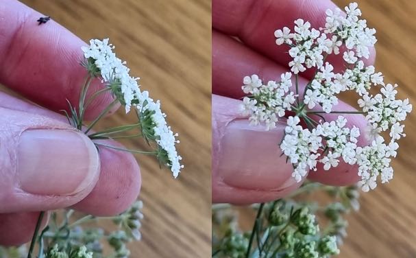
Figure 3. Conium maculatum flowers
Review of Hemlock Poisoning
Hemlock is a widespread biennial herb that commonly grows in undisturbed, moist, or damp areas, including near stockyards, along the banks of rivers or creeks, and along roadsides (Cortinovis & Caloni, 2015). It is distinguished by its fern-like leaves, white flowers, single taproot, and hollow stems that have small, irregular purple spots (Cortinovis & Caloni, 2015). All parts of the plant are toxic when ingested due to the piperidine alkaloids contained within the plant, namely γ-coniceine and coniine (Galey, Holstege & Fisher, 1992). Fresh plant material is thought to have the highest concentrations of γ-coniceine, whereas the seeds and dried plant material are thought to contain mostly coniine (Galey, Holstege & Fisher, 1992). The toxicity of hemlock in dried forage may decrease over time due to the volatility of these poisonous compounds (Galey, Holstege & Fisher, 1992). This instability may also provide reason behind the characteristic, offensive "mousy odour" that is often reported to be smelt on the gastrointestinal contents, breath and urine of affected animals as well as when the leaves of the plant are crushed (Galey, Holstege & Fisher, 1992). This smell was not detected from any of the animals or plants in this case.
The acutely toxic properties of hemlock are well recognised yet there is limited recent literature describing livestock poisonings due to hemlock ingestion. Due to the lack of characteristic pathology, diagnosis is based on exclusion of other potential causes combined with clinical signs and demonstration of accessibility to the plant. Binev, Mitev & Miteva (2007) describe the analysis of urine and forage samples for coniine and γ-coniceine alkaloids, however the practicality of this diagnostic technique may be limited. Hemlock toxicity does not result in any specific haematological changes (Binev, Mitev & Miteva, 2007), which is consistent with the non-specific, mild muscle and hepatic enzyme increases identified during our investigation. Ketosis was a recurring finding in this case and likely secondary to inappetence and reduced feed intake.
Conditions of Poisoning
Piperidine alkaloids are thought to act by stimulating and then paralysing the nicotinic receptors of the central and peripheral nervous system (Galey, Holstege & Fisher, 1992), leading to hypoxia and anoxia of the brain's respiratory centre, which eventually causes respiratory depression and failure, leading to death (Galey, Holstege & Fisher, 1992; Binev, Mitev & Miteva, 2007). Sources suggest fresh hemlock may be lethal to bovines at 5.3g/kg of bodyweight, and coniine lethal at 3.3mg/kg (Binev, Mitev & Miteva, 2007; Vetter, 2004). Cattle are believed to be the most sensitive to the plant, followed by horses, pigs, sheep and finally goats (Binev, Mitev & Miteva, 2007). It could be proposed that the piperidine alkaloids found in hemlock might have an addictive nature when consumed, like related alkaloids such as nicotine, which facilitate the release of neurotransmitters such as dopamine upon nicotinic receptor activation (Furbee, 2009).
Clinical signs of toxicity have been observed 30-40 minutes after ingestion. These signs include nervousness, tremors and ataxia, increased urination and defecation, hyperpnoea, tachycardia, followed by depression and recumbency (Cortinovis & Caloni, 2015). A comatose state progressing to death from respiratory failure may also occur (Cortinovis & Caloni, 2015). Amongst the clinical signs reported was hyperthermia, which was a defining feature of the disease process in this case (Galey, Holstege & Fisher, 1992). It may result from alkaloids promoting peripheral vasoconstriction or depression of the thermoregulatory centre due to hypoxia from alkaloid action (Binev, Mitev & Miteva, 2007). Reports of elevations in liver enzymes are likely due to the hepatotoxic effects of the plant, which is corroborated by the lab findings in our case (Binev, Mitev & Miteva, 2007). Teratogenic effects of hemlock ingestion below the toxic threshold in various species are also described (Galey, Holstege & Fisher, 1992; Binev, Mitev & Miteva, 2007; Vetter, 2004). Affected neonates are born with neurological deficits and developmental abnormalities, including congenital skeletal contractures and cleft palates (Galey, Holstege & Fisher, 1992; Binev, Mitev & Miteva, 2007; Vetter, 2004).
Treatment, Management and Recommendations
There is no known cure for hemlock ingestion and poisoning in livestock (USDA Agricultural Research Service, 2018). Treatment is aimed at removal of the stock from the plant source and providing supportive care (USDA Agricultural Research Service, 2018). It should include provision of shade, adequate fresh drinking water and good nutrition (USDA Agricultural Research Service, 2018). Decontamination via gastric lavage or administration of activated charcoal may be beneficial to prevent further absorption (USDA Agricultural Research Service, 2018). Administration of atropine may temporarily alleviate parasympathetic effects such as increased salivation and lacrimation, however Galey, Holstege & Fisher (1992) reported that this treatment offered no significant improvement for respiration or the course of the disease. Sedatives such as diazepam may be used to control convulsions, muscle fasciculations, and subsequent hyperthermia (McKenzie, 2012). Provision of broad-spectrum antimicrobials and non-steroidal anti-inflammatories may be indicated to prevent secondary aspiration pneumonia (Poulsen, 2022).
Due to the dramatic presentation of hemlock poisoning, it has been suggested that cases of livestock deaths due to hemlock poisoning are either incorrectly attributed to another cause or occur infrequently despite the widespread distribution of the weed (Powell, 1992). Recommendations for future management focus around landholder awareness of the plant and its toxic effects. Weed management principles can be applied to include prevention of spread, mechanical or chemical control of the plant, and grazing management techniques to ensure cattle are not overgrazing paddocks where they would be more likely to ingest the plant (Graham & Johnson, 2004). Western water hemlock, a similar toxic plant, is reported to be more palatable after herbicide treatment (Duggan, 2018). Animals are more likely to consume the plant when there is a lack of alternative forage, particularly if the plant is in a young, active growth stage (Duggan, 2018). Hemlock can also be poisonous if ingested as hay or silage, thus accurate identification of plant species in a pasture is essential prior to fodder preparation (Duggan, 2018). It may be worthwhile to offer training to landholders in plant identification and differentiation between hemlock and similar weeds, to reduce the risk of losing valuable animals, as in this case.
References
- McKenzie RA (2012) 'Part 3 - Poisonous Vascular Plants' in Australia's Poisonous Plants, Fungi and Cyanobacteria, pp 232-234 CSIRO PUBLISHING
- Hepworth K (2018) Thiamine deficiency induced polioencephomalacia (PEM) of sheep and cattle www.agric.wa.gov.au Accessed April 2023
- Daly R (2021) Ergot: A Potential Livestock Poisoning Problem extension.sdstate.edu Accessed April 2023
- Shepherd RCH (2010) 'Thymelaeaceae' in Is That Plant Poisonous?, pp 192-193 RG and FJ Richardson
- Cortinovis C and Caloni F (2015) Alkaloid-Containing Plants Poisonous to Cattle and Horses in Europe Toxins 7(12):5301-5307
- Galey FD, Holstege DM and Fisher EG (1992) Toxicosis in dairy cattle exposed to poison hemlock (Conium maculatum) in hay: isolation of Conium alkaloids in plants, hay, and urine Journal of Veterinary Diagnostic Investigation 4:60-64
- Binev R, Mitev J & Miteva T (2007) Intoxication with poison hemlock (Conium maculatum) in calves Trakia Journal of Sciences 5(3-4):40-50
- Vetter J (2004) Poison hemlock (Conium maculatum L.) Food and Chemical Toxicology 42:1373-1382
- Furbee B (2009) 'Chapter 47 - Neurotoxic Plants' in Clinical Neurotoxicology, pp 523-542 WB Saunders
- USDA Agricultural Research Service (2018) Poison Hemlock (Conium maculatum) www.ars.usda.gov Accessed April 2023
- Poulsen KP (2022) Aspiration Pneumonia in Large Animals www.msdvetmanual.com Accessed April 2023
- Powell JM (1992) Conium maculatum L. plantnet.rbgsyd.nsw.gov.au Accessed April 2023
- Graham J and Johnson W (2004) Managing Poison and Western Water Hemlocks extension.unr.edu Accessed April 2023
- Duggan S (2018) Poison hemlock and Western waterhemlock: deadly plants that may be growing in your pasture extension.oregonstate.edu Accessed April 2023