CASE NOTES
Lumpy Skin Disease Exclusion - A Case Study
J McNally, District Veterinarian, North West Local Land Services, Moree
Posted Flock and Herd July 2023
Following the detection of Lumpy Skin Disease (LSD) in cattle in Indonesia in March 20222, the Australian veterinary authorities advised veterinarians and participants in the livestock industry to have a heightened awareness for clinical signs in cattle and buffalo that were consistent with LSD. The reason for this heightened awareness was the increased threat of an LSD incursion in Australia as the disease was now present in a neighbouring country and had been rapidly moving through south Asia for the past two years2.
Lumpy Skin Disease is a highly contagious viral disease of cattle and buffalo1, the LSD virus being a species within the Capripoxvirus genus. Whilst it does not have a high mortality rate it can result in animal welfare issues and production losses1. It would also have a significant impact on our domestic and international trade, with Australia no longer being recognised as free from LSD1. The economic losses to Australian livestock producers and the associated industries would be significant and ongoing1.
The principal means of transmission of LSD is by arthropod vectors, such as biting flies, mosquitoes and ticks(1,3). It is also believed that there may be some transmission by direct contact between animals(1,3). The disease can be spread over long distances via the movement of infected animals or contaminated equipment, vehicles or the clothing and footwear of people1.
In light of this increased potential of an LSD incursion, thoroughly investigating any case that could include LSD as a differential diagnosis was a priority. In March 2022 I received a call from the EAD Hotline on-call veterinarian regarding a potential LSD case that was reported in the Moree district. Moree is situated in the north-western area of New South Wales on the Queensland border and is one of the most agriculturally productive local government areas in Australia, with a Gross Regional Product of more than $750 million(4). The case in question was a heifer in a feedlot 60km north-west of town. The feedlot staff had noticed multiple, raised skin lesions across the beast during a routine weighing procedure.
A visit was organised for the following morning and preparations were made to ensure a thorough investigation could be conducted swiftly. Photos were sent through from the feedlot manager which allowed an initial assessment of the beast, and from these photos LSD was high on the differential diagnosis list (see Figure 1 and Figure 2). A good history was obtained over the phone from the feedlot manager to ascertain if there were significant risk factors that could indicate LSD was likely. Contact was made with the senior government veterinarian in the NW, as well as EMAI to ensure they knew an LSD exclusion was being sent in the next 24 hours.

Figure 1. Photo of affected beast sent by feedlot manager

Figure 2. Photo of traumatised lesions sent by feedlot manager
Prior to arrival at the feedlot I had also refreshed my knowledge on the epidemiology of LSD. The virus is spread largely through arthropod vectors and less commonly through direct contact. Being a feedlot and the concurrent environmental conditions in the district, spread to other cattle in the feedlot was a likely scenario. LSD lesions can take between 4-20 days to develop following 'inoculation', thus it would be very important to establish when this beast arrived on the property. The disease has a morbidity of 5-45%, so if this was a case of LSD there could potentially be more affected animals at the feedlot.
On arrival at the feedlot the following morning, I ran through the known history of the beast to try an establish a risk grading. The heifer was transported to the feedlot from Dalby Saleyards 41 days prior to lesions being seen. During the 41 days on feed no lesions had been noted on this animal until the day prior to my visit, and no lesions had been seen on other cattle in the feedlot. Staff did note that the heifer was a more aggressive animal than most of the other cattle but had been gaining weight whilst on feed. The heifer was pulled the day before my visit, as a pen-rider had noticed she looked depressed and from a distance her coat looked rough. On examination in the crush, staff saw multiple, raised lesions over the entire body and head of the heifer. They immediately called their feedlot veterinarian who instructed them to call the EAD Hotline at once.
Following discussions with the feedlot manager I examined the heifer in the crush. She had multiple, firm, raised, hair-covered lesions across her face, torso, neck, legs, anus, vulva, tail, edge of eyelid and conjunctiva (see Figures 3, 4 and 5). They were of various sizes but ranged from approximately 3mm to 25mm, with some coalescing due to the sheer number of lesions. Some lesions were bloody due to trauma but did not appear to be ulcerated lesions. The heifer's rectal temperature was 40.7°C. She had increased clear nasal, ocular discharge and increased salivation. She was also agitated.

Figure 3. Lesions on clinical examination
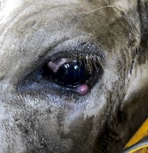
Figure 4. Eye lesion

Figure 5. Eye lesion
From gross examination the lesions were similar to those described in LSD cases but the lack of ulceration of lesions and the sheer number of lesions made LSD a less likely diagnosis. An area of hair was also shaved to further assess the lesions and they appeared in be within the subcutaneous layer (see Figure 6). Biopsies were taken and grossly the tissue appeared to resemble lymphoid tissue. Fine needle aspirates were also performed on two lesions; scabs were removed from two lesions - one placed in PBSG and one in a dry sterile container; bloods were also taken.

Figure 6. Shaved area on side of beast
Following the visit and collection of samples, my differential diagnosis list included LSD, Pseudo cowpox, Bovine herpes virus 2 and an allergic reaction. I also personally thought the tissue biopsied looked like lymphoid tissue and I wondered if this could be a case of lymphoma or lymphosarcoma. All samples were sent to EMAI following their EAD submission protocol.
Results from AAHL and EMAI confirmed negative results for LSD, Capripoxvirus (LSD), Herpesvirus, Parapoxvirus, Enzootic Bovine Leucosis, and Pestivirus. Electron microscopy was also negative for any virus. Histopathology examination concluded that the lesions were the result of a Round Cell neoplasm, with lymphoma being favoured.
Six days later I was called back to the feedlot as the heifer had further deteriorated. The heifer was now laterally recumbent and unable to support herself on her brisket or rise. When approached she showed signs of altered mentation, being agitated and aggressive, and began to paddle. The decision was made to euthanase her and perform a full post-mortem.
On post-mortem the extent of the distribution of the lesions was remarkable. The lesions were numerous, discrete, firm, cream to light pink and red to purple, spherical and oval lesions throughout every tissue plane and most organs. Some lesions were solitary, whilst others were in conglomerations. When palpated the lesions were firm, with a consistent consistency and colour when incised (see Figures 7, 8, 9, 10, 11, 12 & 13).

Figure 7. Muscle Layer

Fgure 8. Subcutaneous layer

Figure 9. Heart
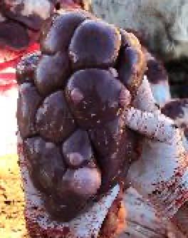
Figure 10. Kidney

Figure 11. Kidney

Figure 12. Muscle Layer

Figure 13. Sinus
In the thoracic cavity there were lesions embedded in the musculature and a number of small lesions were 'free-floating', but there were no lesions within the lung tissue. The heart was also enlarged and there was a pericardial effusion and evidence of fibrin attachments between the pericardium and the serosa of the heart. The heart muscle appeared grey in colour and there were white striations on the surface. A lesion was found near the apex and a dark red lesion found on each of the mitral and tricuspid valves.
Both the spleen and the kidneys had lesions throughout them but the liver did not appear to have any lesions. In the kidneys the lesions extended from the capsule to the cortex and were well demarcated.
The submandibular lymph nodes were enlarged and cream to grey in colour with a mottled appearance on the cut surface. There were also lesions seen on the mucosal surface of the sinus.
Most interestingly, the dura mater was thickened and very difficult to cut. When the brain was removed the dura mater appeared to be of variable thickness with an irregular surface which was pink on the inner aspect (see Figure 14).

Figure 14. Dura mater
Samples were again sent to EMAI for histopathological examination and to determine the classification of the round cell neoplasia. The final diagnosis was Lymphoma of T cell origin, and this was determined by immunohistochemistry. In conclusion the beast suffered from Sporadic Bovine Leucosis.
References:
- The facts of Lumpy Skin Disease www.agriculture.gov.au April 2023
- Lumpy Skin Disease transmission www.dpi.nsw.gov.au April 2023
- Lumpy Skin Disease FAQ www.woah.org April 2023
- Overview of Moree Plains Shire Council www.mpsc.nsw.gov.au April 2023