Multiple Presenting Syndromes as a Result of Bovine Anaemia Caused by Theileria orientalis Group
Andrew Biddle, District Veterinarian, New England LHPA
Posted Flock & Herd September 2012
SUMMARY
In March 2012 significant losses were incurred on two neighbouring properties as a result of infection with Theileria orientalis group. On the first property a group of 12 cows and two bulls were affected and on the second, two cows died and two calves were still born. Both properties had introduced cattle from Southern NSW or Victoria in the preceding three to four months.
History
Both properties are located approximately 45 kms east of Glen Innes. The properties are adjoining but are run and managed independently. Seasonal conditions have been favourable throughout the summer of 2011/12. Pasture quality and quantity was excellent.
Property 1 was part of a Theileria prevalence survey undertaken in 2010. At this time 10 cows were blood sampled and smears made for microscopy to look for Theileria spp. Organisms were only found in one of ten high power fields from one in ten animals sampled. No previous testing had been undertaken on property Two.
Both properties had introduced the affected groups of animals from Southern NSW or Victoria in the preceding three months.
At Property 1, cattle were purchased at a stud dispersal sale near Albury in December 2011.
At Property 2, cattle were purchased from Southwest Victoria at a similar time.
Clinical Signs
The first visit to Property 1 was on the 14th March following a request by the owners to examine a group of cows and two bulls that had been losing weight and were generally of illthrifty appearance. Other cattle on the property were fat and appeared healthy.
One mature cow, purchased in calf, was pregnancy tested and found to be empty. Five cows and two bulls were sampled.
On the 15th March the neighbouring property (Property 2) reported two ‘still born’ calves. These two calves were necropsied and the cows were blood sampled.
Post-mortem findings
The two dead calves examined on Property 2 were ‘stillborn’ as neither had suckled, walked or taken a breath. The calves were fully developed and full term.
Calf 1: The calf had a pleural effusion, straw coloured abomasal and engorged blood vessels on the meningeal surface of the brain.
Calf 2: The calf had a mottled appearance to the liver, and a serosanguinous pleural effusion in the abomasum. The abomasal fluid was dark in colour.
On the 19th of March I revisited Property 2 to post-mortem the mother of calf 2 and another cow that had been found dead overnight (Figure 1).

Figure 1.Cow found dead overnight

Figure 2. pale and yellow mucous membranes
There were no signs of kicking or struggling prior to death and the ocular mucous membranes were pale and yellowish (See Figure 2).
Subcutaneous and abdominal fat was yellow and the liver was enlarged and ochre coloured (See Figure 3).

Figure 3. Enlarged ochre liver
Blood that pooled after the foreleg was reflected was semi translucent and did not clot (see Figure 4).
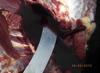
Figure 4. The blade of the knife can be seen through the blood that appears more like raspberry cordial.
Laboratory results
Property 1:
| Identity | PCV | Smears | Comments |
|---|---|---|---|
| Cow 1 | 13 | Theileria in 2.5% of RBCs | |
| Cow 2 | 18 | Theileria in 3.1% of RBCs | Regenerative anaemia |
| Cow 3 | 29 | Theileria in 3.3% of RBCs | |
| Bull 1 | 40 | Theileria in 1.0% of RBCs | |
| Bull 2 | 39 | Theileria in <1 % of RBCs |
Property 2:
| Identity | PCV | Smears | Comments |
|---|---|---|---|
| C1769 | 15 | Theileria in 40 % of RBCs | Regenerative anaemia |
| A1186 | 16 | Theileria in 35 % of RBCs | Regenerative anaemia |
Theileria major piroplasm surface protein PCR was carried out on the blood samples collected from both properties. The animals were positive for Ikeda, Chitose and Buffeli types.
Discussion
This case illustrates the classic presentation of bovine anaemia caused by Theileria orientalis group whereby naive animals are introduced to an endemic area with carrier animals and tick activity. Differential diagnoses in this case ranged from pestivirus and leptospirosis, as potential causes of still births, through to Anaplasmosis and Babesiosis as the cause of anaemia and jaundice.
In a prevalence survey undertaken in the New England LHPA 70% of herds were found to have Theileria spp. in blood smears (Biddle et al. 2010, unpublished data). No PCR testing was undertaken to determine the sub types. Sporadic clinical cases have been reported in the New England LHPA. These cases have predominantly been in the eastern areas of the Authority.
The disease syndrome associated with Theileria orientalis in NSW has only been recently described (Izzo et al. 2010). It is possible that further clinical cases have occurred in the New England LHPA region. For example, in the past similar cases involving mortalities in recently introduced animals have been investigated. Presenting with jaundice, these have been diagnosed as suspect Bracken Fern toxicity.
On Property 1 the difference between the cows and bulls is worth comment. The bulls were kept near the house in cleared paddocks at least one km from timbered areas. The cows were in paddocks that contained timbered areas and were mixed with home bred animals. The bulls were clinically affected (ill thrifty) but it was the cow group where the greater clinical effects (anaemia and abortion) were found. The cows also had a greater parasitic burden. In the paddocks where the cows grazed there were more likely to be vectors and home bred carrier animals.
The owner of Property 1 indicated that 2011/2012 had seen minimal Buffalo Fly activity compared with previous years. As a result no buffalo fly suppression activities had been undertaken. The property normally uses either organophosphate or synthetic pyrethroid products which although not registered for ticks would likely have suppressed their activity.
References
- Biddle A, Eastwood S, Martin, Freeman 2010. Unpublished data on file NELHPA
- Izzo, M., Poe, I., Horadagoda, N., De Vos, A. and House, J., Haemolytic anaemia in cattle in NSW associated with Theileria infections. Australian Veterinary Journal 2010; 88:45-53