FACIAL ECZEMA IN CATTLE—FMD EXCLUDED
Kylie Greentree, District Veterinarian, Cumberland LHPA
Posted Flock & Herd December 2011
Introduction
This case study outlines a disease investigation that was in response to a farmer contacting the Emergency Disease hotline. The syndrome reported by the farmer could have been consistent with an exotic disease. The case study outlines the approach that the Cumberland LHPA took to reach a diagnosis and exclude important exotic disease.
Figure 1: Cattle with facial eczema constantly licking their noses

History
In early March 2011 the Cumberland LHPA was tasked to respond to a producer enquiry that had come through the Emergency Animal Disease Hotline. A local producer reported three Murray Grey cows with excessive head shaking, bleeding from the nose and frothing at the mouth. When further history was collected on the following day by the Cumberland LHPA, there were now five affected animals and some of the cattle were reported to have a dry cough. It was also reported that 1-2 weeks prior they had had very watery scours.
There were 10 cows in this affected mob. The cows were in a predominant rye grass paddock (also with phalaris and clover) that had been slashed 3 weeks prior. The paddock had been fertilised with chicken manure 12 months earlier. All cows were vaccinated with either 7-in-1 or a 5-in-1, there was a selenium lick block in the paddock and the water (in a concrete trough) looked clear. The cattle ranged in age from 4-7 years. Three cows had calves and did not appear to be affected. All cattle had been moved to an adjoining paddock with similar pasture content but had not been slashed. All cattle were in good condition body score. One cow had aborted a calf and several others that were supposed to be heavily in calf appeared empty.
WEATHER CONDITIONS LEADING UP TO THE EVENT
FEBRUARY
| DATE | MIN-MAX TEMP | RAINFALL | RELATIVE HUMIDITY |
|---|---|---|---|
| 1 | 18.5-38.6 | 0 | 51 |
| 2 | 25.7-35.8 | 0 | 52 |
| 3 | 25.2-37.7 | 0 | 58 |
| 4 | 20.9-34.8 | 0 | 73 |
| 5 | 23.3-38.9 | 0 | 52 |
| 6 | 25.2-34.0 | 0 | 56 |
| 7 | 13.5-20.2 | 11.8 | 93 |
| 8 | 15.0-24.7 | 0 | 84 |
| 9 | 15.1-24.3 | 1.0 | 81 |
| 10 | 16.3-26.7 | 1.2 | 91 |
| 11 | 16.7-34.2 | 0 | 77 |
| 12 | 21.3-22.0 | 0.2 | 90 |
| 13 | 17.3-21.1 | 7.0 | 97 |
| 14 | 16.2-20.2 | 9.8 | 99 |
| 15 | 15.4-22.5 | 0.2 | 96 |
| 16 | 16.4-24.5 | 1.2 | 90 |
| 17 | 18.6-29.7 | 0 | 96 |
| 18 | 19.2-25.3 | 2.6 | 95 |
| 19 | 19.0-34.5 | 0 | 97 |
| 20 | 21.3-34.9 | 0.2 | 64 |
| 21 | 17.8-23.9 | 3.0 | 97 |
| 22 | 14.4-19.9 | 0.8 | 78 |
| 23 | 14.4-22.8 | 1.2 | 92 |
| 24 | 12.0-26.5 | 0 | 92 |
| 25 | 13.1-25.8 | 0.2 | 79 |
| 26 | 15.4-30.3 | 0 | 97 |
| 27 | 16.8-28.7 | 0 | 76 |
| 28 | 19.4-29.6 | 0.2 | 87 |
The mean statistics for February were:
Min-max temp= 18-28.3
Relative humidity= 81%
MARCH
| DATE | MIN-MAX TEMP °C | RAINFALL mm | RELATIVE HUMIDITY % |
|---|---|---|---|
| 1 | 21-32.1 | 5.4 | 61 |
| 2 | 16.1-19.2 | 0.8 | 76 |
| 3 | 15.6-30.4 | 0.4 | 95 |
| 4 | 16.4-29.2 | 0.2 | 81 |
| 5 | 14.9-20.7 | 0.6 | 84 |
| 6 | 10.8-21 | 0.2 | 58 |
| 7 | 13.7-23 | 1.4 | 97 |
Over the entire month of March the mean statistics for the month were:
Min-max temp= 15.9-25.4
Relative Humidity= 86%
The weather leading up to the disease event was favourable for the production of fungal spores. Overnight temperatures were greater than 12°C most nights, there were consistent small amounts of rain to keep the grass moist and the relative humidity during February and March was quite high, sometimes reaching 99%.
Clinical examination
The cattle were very agitated; they were shaking their heads like a dog with a grass seed in its external ear canal. Their noses appeared sunburnt, red and bleeding. The cattle were continuously licking their noses and hypersalivating.
Figure 2: Photosensitisation to the nose

On examination of the tongue, most of the cases had ulceration on the ventral and lateral margins. Several cows had crusty scabs over the skin on the neck and behind the ears. The margins of the eyelids and the vulva appeared crusty and burnt. Only the light coloured Murray Grey cattle were showing these clinical signs. All the cattle that were examined had an elevated temperature 39.4 - 40.5°C but they were also very agitated getting into the crush which would have elevated their temperature.
DIAGNOSTIC AIDS
Serum and EDTA blood was collected from five cattle. These samples were submitted to the State Diagnostic Laboratory to exclude Foot and Mouth Disease, Vesicular Stomatitis and undertake a liver profile.
Results of the liver profiles are summarised in Table One: Liver profiles of serum samples 1-5.
| Sample | 1 | 2 | 3 | 4 | 5 |
|---|---|---|---|---|---|
| GGT ( 0-35U/L) | 1055 | 599 | 734 | 1688 | 296 |
| GLDH (0-30U/L) | 1595 | 759 | 472 | 657 | 87 |
| AST (0-120U/L) | 478 | 129 | 340 | 155 | 294 |
| BIL (0.0-24.0umol/L) | 38.8 | 138.9 | 116.4 | 48.2 | 148.1 |
| BIL-C umol/L | 23.8 | 64.4 | 71.2 | 21.8 | 35.7 |
| CK (0-300U/L) | 752 | 156 | 385 | 183 | 977 |
| PROTEIN (60-85g/L) | 71.8 | 73.9 | 69.6 | 81.8 | 70.3 |
| ALBUMIN (25-38g/L) | 32.6 | 34.7 | 39.5 | 33.9 | 33.4 |
| GLOB (30-45g/L)v | 39.2 | 39.2 | 30.1 | 47.9 | 36.0 |
| ALB/GLOB (0.7-1.1) | 0.8 | 0.9 | 1.3 | 0.7 | 0.9 |
The liver profile results were interpreted by a pathologist from the EMAI State Diagnostic Laboratory as being indicative of hepatopathy with biliary disease. Elevated CK and AST are likely due to myopathy associated with recumbency.
Figure 3: Predominant Ryegrass pastures

The PCR results for Foot and Mouth and Vesicular Stomatitis were negative
Pasture samples were taken from two paddocks and also submitted to the State Diagnostic Laboratory:
1. The initial Ryegrass paddock that had been slashed and the Ryegrass was lying in wind rows.
2. The second paddock that they went into that was not slashed but was still predominant Ryegrass.
The report from the laboratory indicated that there was an average of Pithomyces at 175, 000 spores per gram of pasture and that this level is dangerous. Interpretation also included the comment that counts of 100, 000 will cause facial eczema and photosensitisation. Higher counts such as seen here will almost certainly cause liver damage in grazing sheep and cattle. This is also indicated by high serum levels in the liver of the affected animals. Parkinson, Malmo and Vermunt (2010) also report that counts of 40,000 spores per gram of pasture over many days can also be toxic especially when animals are grazing towards the base of the sward.
Both paddocks had a similar spore count so it was recommended that the few cows and calves in the second paddock were removed from this paddock also.
On the 28th March 2011 the producer took further pasture samples from the two paddocks in question. Over the 3 weeks there had been some considerable rain (71.4mm). The results on these paddocks were:
Sample 1: (Upper Paddock): 85,000 spores/gm pasture
Sample 2: 100,000 spores/gm pasture
In May 2011 the property was revisited by the Cumberland LHPA as another cow had lost a lot of condition over the last two weeks. On clinical examination of the cow you could see that she had skin damage to the nose that had recovered and that there was a decrease in the pigmentation. Bloods were taken and a liver profile requested. The results are summarised in Table Two:
Table Two: Laboratory results for bloods collected in May 2011.
| Lab parameter | Result |
|---|---|
| GGT 0-35 U/L | 1123 H |
| GLDH 0-30 U/L | 62 H |
| AST 0-120 U/L | 142 H |
| BIL 0.0-24.0 umol/L | 17.3 |
| BIL 0.0-24.0 umol/L | 17.3 |
| CK 0-300 U/L | 496 H |
| PROTEIN 60.0-85.0 g/L | 72.3 |
| ALBUMIN 25.0-38.0 g/L | 32.7 |
| GLOB 30.0-45.0 g/L | 39.6 |
| ALB/GLOB 0.7-1.1 | 0.8 |
The liver profile indicating a severe cholangiohepatopathy.
Parkinson, Vermunt and Malmo (2010) indicate that serum GGT gives the best indication as to the severity of bile duct damage. A serum concentration of GGT between 70-300IU/L may be seen with mild liver damage, 300-700IU/L with moderate damage and higher than 800IU/L with severe liver damage. GGT levels between 30-70IU/L are not clinically significant.
Figure 4: Ulceration of the tip of the tongue - Foot and Mouth exclusion was done

Post-mortem
No post-mortems were undertaken. One cow died but was buried immediately. As the owner knew the cause of death she did not ring for a post mortem to be done.
If a post-mortem was done on these cattle you would expect:
Macroscopic evidence of oedema and fibrosis of the biliary tracts
Histologically: duct hyperplasia and fibrosis may be seen
Chronic cholangitis will eventually lead to left lobe atrophy in the liver of ruminants. Cattle with an enlarged right lobe and a thin, tough and small left lobe should always consider the possibility of sporidesmin toxicity.
Diagnosis
A diagnosis of Facial Eczema was made on the basis of the history, clinical signs, liver profile and fungal spore results.
Follow up
Over the few months since the initial incident of Facial Eczema there have been two deaths. One died 3-4 days after the first visit on the 7th March 2011 and the other cow died mid June 2011. The first one that died only had moderately high GGT levels but was very agitated on examination, and the other cow (Z6) was not blood tested. ‘Z6’ lost weight over several months and was found dead. The producer said she definitely had the same clinical signs as the other Facial Eczema cases.
Three months after the initial visit the remaining cows have put back on some condition and seem to be doing well. The last cow we blood tested appears bright and eating well but is still very thin so it will be interesting to see how she progresses.
Discussion
Facial Eczema is caused by the ingestion of fungal spores of Pithomyces chartarum. These spores contain the hepatotoxic compound, sporidesmin. The liver damage from sporidesmin causes the photosensitisation lesions that are a characteristic symptom of facial eczema. Facial Eczema normally occurs in hot humid conditions, in late summer to early autumn. The greatest risk occurs when humidity is close to 100% and when the grass temperatures have reached a minimum of 12°C for 4 consecutive nights. These conditions are normally found when more than 4mm of rain falls within 48hrs or when soil is kept moist by irrigation. Two or more repetitions in these weather conditions can lead to the fungus increasing to dangerous levels.
The fungus grows on dead litter of pasture, but can be found along the entire sward. The disease tends to be associated mainly with Perennial Ryegrass (Lolium perenne) because of its ability to produce dead litter.
When the spores are eaten by the ruminant, the spores release the sporidesmin toxin in the digestive tract. From here it is absorbed into the bloodstream, removed by the liver and excreted into the biliary system. The sporidesmin concentrates in the biliary system causing severe necrosis of the mucosal surface of the biliary ducts. The resulting peri-cholangitis leads to obstructive jaundice and accumulation of phytoporphyrin in the skin. The reaction between sunlight and circulating phytoporphyrin leads to tissue damage especially at muco-cutaneous junctions in lightly coloured or non-pigmented skin. Dark pigmented cattle may not show clinical signs.
Facial eczema can affect all ruminants to varying degrees. Alpacas and sheep are highly susceptible, cattle are moderately susceptible and goats have low susceptibility. Interestingly some family lines have greater susceptibilities, which illustrate the importance of selecting for resistance.
When the investigating veterinarian collected the initial history and heard that the cattle were head shaking they developed a list of differentials focused on neurological head shaking:
- Ryegrass staggers (considered the most likely given that we were on a Ryegrass paddock and the time of year)
- Polioencephalomalacia (normally associated with thiamine deficiency, or high sulphur intake)
- Lead poisoning (no known lead source in the paddock)
- Botulism (had used chicken manure but 12 months before and never put stock on paddocks directly after application and did quarantine paddocks but was still considered possible)
- Nervous Ketosis (protein and energy insufficiency was a possibility given that the cows were late stage pregnancy but is more common during early lactation)
- Urea poisoning (unlikely as no urea being fed in a supplement and no sudden death)
- Pituitary abscess (uncommon and normally individual cases, often associated with putting a nose ring in a bull)
- Thromboembolicmeningoencechalitis (highly unlikely normally seen in young feedlot cattle caused by Histophilus somni)
- Listeriosis (unlikely usually associated with feeding poor-quality silage)
- Once further history was collected the investigating veterinarian considered that secondary photosensitisation was more likely. Photosensitisation can be classified into:
- Primary photosensitisation (photodynamic agent is ingested or injected associated with a variety of photodynamic agents)
- St Johns Wort (Hypericum perforatum)
- Some species of ryegrass, clovers, Lucerne, burr trefoil and Brassica may cause primary photosensitisation
- Drugs such as phenothiazine, tetracycline some sulphonamides and corticosteroids
- Hepatogenous photosensitisation (secondary photosensitisation associated only with phytoporphyrin as photodynamic agent)
- Facial eczema
- Blue-green algae
- Some plants; Panicum, Lantana and Crotalaria species, lupins, ragwort, Ngaio and Caltrops
- Liver fluke or liver abscesses
- Leptospirosis
- Congenital Photosensitisation
- Congenital porphyria
- Photosensitisation of unknown aetiology
- Weaner calves (chlorophyll overload and an underdeveloped liver which is unable to cope with the phytoporphyrin) Spring eczema.
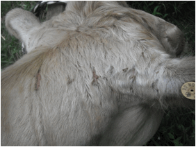
Figure 5: Scabbing of the skin on the neck and behind the ears
Mortalities are generally not very high with Facial Eczema however severely affected animals may have many problems in following seasons. These problems include metabolic problems related to calving and fertility or further photosensitisation. Even though the liver regenerates it may be enough to cope with their normal daily requirements but not enough to cope with a growing foetus. In general young stock recovers better than older stock.
Treatment
There is no specific treatment for Facial eczema - any treatment should be symptomatic.
Shelter is essential, allowing the current lesions to heal and no further skin lesions to occur. The property these cattle were on fortunately had a lot of trees and the cattle were seeking the shade anyway.
I also recommended sun-block for the noses and other areas where skin was peeling off these cattle, but it was an impossible task especially with the cattle being so agitated. These cattle were show cattle and normally very friendly but the diseased cattle were very stressed.
Anti-inflammatories were recommended to help with the discomfort but once again they were too difficult to get into the yards so we thought it was better to leave them alone.
Antibiotics if skin lesions become infected
Good quality diet.
Figure 6: Photosensitisation to the nose, crusting around the eyes and hyper salivation

Prevention
- Predicting dangerous periods based on pasture content and weather patterns
- Spore counting of pastures (Certain areas have higher spore counts; north facing slopes and ridges and ground near hedges are areas associated with higher spore counts)
- Avoid hard grazing of paddocks when these weather conditions are suitable, move cattle to the longest pasture possible
- Avoid paddocks cut for hay or late topped, they are more likely to be more toxic because of greater amount of pasture litter
- In general paddocks that are sheltered by wind breaks or hills are more dangerous
- It is believed that the warmer northern slopes carry higher risk than the cooler southern slopes
- Summer growing crops are generally safer than pastures
- Provide alternative feed during these danger periods
- Minimise the build up of dead matter
- Monitoring spore counts; faecal spore counts is a relatively new method of monitoring exposure to Pithomyces spores. (not known to be done in Australia)
- Spraying high-risk farms with a fungicide (This is commonly done in New Zealand but not economically feasible in Australia) Fungicides are ineffective with spore counts greater than 200000/g grass, and the effectiveness of some sprays will decrease with more than 25mm of rain
- Dosing animals with zinc 2 to 3 weeks before the danger period can prevent the deleterious effects of sporidesmin on the liver ZnSO4 in water (25-30g/cow), ZnO in drench, in feed or on pasture or as intraruminal capsules (not available in Australia). Dosing with Zinc after the effects of sporidesmin have occurred will not decrease the liver toxicity.
- Selecting for resistant cattle; select against high GGT concentrations following Facial eczema.
References
- Diseases of Cattle in Australasia TJ Parkinson, JJ Vermunt and J Malmo
- www.weatherzone.com.au