CASE NOTES
LUNG WORM INFESTATION IN NORTH COAST BEEF WEANERS
Ainslie Lund, North Coast Livestock Health and Pest Authority, Grafton, NSW
Posted Flock & Herd May 2014
History
In August 2013 The North Coast Livestock Health and Pest Authority (NCLHPA) was called to investigate poor growth, diarrhoea and death in a group of 15 beef weaners near Grafton, NSW. The weaners ranged in age from 6-8 months. The weaner group contained a mix of breeds and their crosses including Angus, Murray Grey, Jersey and some with Bos indicus infusion. The farmer reported that the group had showed ill thrift for a number of months. The cattle were weaned early as many lost their mothers in the January 2013 floods. The weaners were moved to the current paddock 3 months prior to the NCLHPA visit. They had not been drenched in this time and the farmer was unsure if they had ever been drenched. The pasture was native and unimproved with large areas of swamp and marsh. There was ample dry standing feed but feed quality was poor. This paddock was recently purchased by the current owner. Its grazing history prior to these weaners was unknown.
Approximately a week prior to the NCLHPA visit 2 weaners died suddenly with a short illness of 2 days. The day before the NCLHPA visit, 1 heifer had died and on the day of the visit a steer was down. All animals showed signs of weight loss and scours prior to death. The owner had noticed some animals, but not all, had a cough. The owner had suspected flood mud scours (yersiniosis) and administered oxytetracycline (Alamycin 300 LA) to all scouring animals, with no improvement seen.
Clinical examination
The surviving group of animals were viewed in the paddock. All had stunted growth and rough dry coats. At least 4 of the animals were clearly dyspnoeic. They had their necks extended, tongues protruding and were open-mouth breathing. The down steer was in poor body condition with a body condition score of 1.5/5. It was in sternal recumbency but unable to rise. The steer was humanely euthanased.
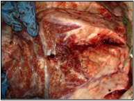
NECROPSY
A post-mortem was performed on the down steer. Significant findings included pallor of the entire carcass (Image 1). The lungs did not deflate when the thoracic cavity was opened (Image 2). Further exploration of the lungs revealed a significant Dictyocaulus viviparus (large lungworm) burden in the terminal bronchioles and lung parenchyma (Images 3 and 4). There was copious amounts of mucus and foam in the bronchioles. There was a significant and clearly visible Haemonchus contortus burden in the abomasum. The wall of the abomasum was oedematous and red.
Image 2: Thoracic cavity

Image 1: Pale carcass

Image 3: Dictyocaulus viviparous

Image 3: Dictyocaulus viviparous
Diagnosis
A diagnosis of internal parasitism, Dictyocaulus viviparous and Haemonchus contortus, was made based on gross evidence at post-mortem.
Discussion
Dictyocaulus viviparus (large lungworm) is a threadlike, creamy white parasite reaching up to 7.5cm in length (Hungerford 1990). Adults live mainly in the bronchi of cattle (Love and Hutchinson 2003), but can extend into the trachea in heavy infections (Hungerford 1990). Female worms produce eggs containing fully developed larvae, which hatch almost immediately. The L1 larvae migrate up the trachea, are swallowed and pass out in the faeces. Development into the infective L3 stage can occur within 5 days under optimal conditions (Taylor et al. 2007). L3 larvae may persist for over 12 months in moist conditions with moderate temperatures of 18°C-21°C (Vermunt et al. 2010). The L3 reach the pasture from the faecal pat either by their own motility or through the ubiquitous fungus agent, Pilobolus (Taylor et al. 2007). Transmission occurs when cattle ingest L3 while grazing (Vermunt et al. 2010).
The disease is almost entirely confined to grazing cattle (Vermunt et al. 2010), with young animals being the most susceptible group. However, the development of highly effective anthelmintic strategies has resulted in dramatic improvements in worm control in first year grazing cattle, and clinical disease is now seen much less commonly (Vermunt et al. 2010). Clinical cases can still occur in immunocompromised or naive animals when sub-optimal stock management is combined with favourable weather conditions and larval pasture contamination.
The climatic conditions on the North Coast favour the survival of all parasitic eggs and larvae for prolonged periods of time. In this specific case, to an unknown extent the pasture was contaminated with lung worm larvae. The young weaners placed on this pasture were very susceptible to disease based on the fact that they were weaned at a young age and not provided with adequate nutrition at any point throughout their life. There was no drenching program in place for this group of cattle. This resulted in an additional high Haemonchus contortus burden leading to anaemia, indicated by the marked pallor of the carcass. The blood and protein loss resulting from the significant Haemonchus contortus burden would of further contributed to their poor health and immunocomprimised state.. These environmental, host and management factors were all contributing factors in the outbreak of lungworm in these animals.
It was recommended that all cattle in this group be treated with a macrocyclic lactone drench with a registered claim against lungworm and Haemonchus contortus. The weaners should be moved to a less contaminated pasture and provided with good quality roughagead lib if pasture quality and quantity is not sufficient. If this paddock must be used it should be grazed with healthy adult cattle. The farmer was recommended to consider the trial use of a drench to all unweaned young cattle at approximately four months of age to ensure this problem did not occur again, or at the very least a drench should be applied to the young animals that will be grazing this particular paddock in the future. Drenching with product/s with more than one active ingredient should occur to delay the onset of parasite resistance.
References
- Hungerford (1990) Diseases of Livestock 9th Edition. McGraw-Hill Book Company Australia Pty Limited
- Love SCJ and Hutchinson GW (2003) Pathology and diagnosis of internal parasites of ruminants in Gross Pathology of Ruminants, Proceedings 350, Post Graduate Foundation in Veterinary Science, University of Sydney, Ch. 16, pp. 309-38
- Taylor MA, Coop RL and Wall RL (2007) Veterinary Parasitology 3rd edition. Blackwell Publishing
- Vermunt JJ, Malmo J and TJ Parkinson (2010) Diseases of Cattle in Australia. Chapter 5 Respiratory Conditions. Vet Learn, New Zealand