
CASE NOTES
Vitamin A deficiency in backyard poultry in north-western NSW
Justine McNally, District Veterinarian, Moree
INTRODUCTION
Nutritional deficiencies occur when poultry are fed a diet deficient in a certain element or when there is inadequate consumption of that element. Vitamin A deficiency in poultry is rarely seen in the commercial poultry industry as complete vitamin premixes are added to domestic poultry diets. Vitamin A is often supplied as retinol, in the form of retinyl acetate, in these premixes and therefore prevents the occurrence of acute hypovitaminosis A1. In backyard poultry operations the risk of hypovitaminosis A increases due to inappropriate management decisions. Common causes are owners developing their own feed mixes without consideration to trace elements, failure to allow birds to access green feed and mishandling during storage of premixes leading to degradation of the product.
OVERVIEW
Vitamin A is physiologically essential for the maintenance of poultry health and ensuring productivity.1,2 Vitamin A is a fat-soluble vitamin and can only be obtained through the diet.2 There are three forms of vitamin A - retinol, retinal and retinoic acid.1,2 It is an essential vitamin for eyesight, maintenance of epithelial and mucosal surfaces, reproduction, embryonic development, growth and maintenance of the immune system.1,2
Vitamin A can be found in both animal-derived feeds such as fish liver oil and fish meal1, as well as plants. Plants are not a good direct source of vitamin A, but a variety contain pigments called carotenoids1 that are converted to vitamin A via metabolic conversion.1,4 In poultry diets the main sources of natural carotenoids are corn gluten meal, yellow corn, algae meal and lucerne meal.2 Conversion of carotenoids to vitamin A can be affected by high environmental temperatures, viral infections, other dietary imbalances and other stressful events.1,2 Gastrointestinal disorders such as parasitism and coccidiosis can also affect absorption of vitamin A due to intestinal wall injury.1,2
Vitamin A is stored in the liver of poultry and thus adult birds can compensate for a vitamin A deficient diet for 2-5 months.3 Chicks acquire vitamin A from their dam and therefore signs of deficiency depend on the quantity of vitamin A passed from the breeder hen. If the breeder hen provided a good reserve of maternal vitamin A, a chick on a deficient vitamin A diet may not show signs for up to seven weeks.3 A chick that hatches with low vitamin A stores may present with signs of hypovitaminosis A during the first week of life.3
There are a range of clinical signs seen with hypovitaminosis A in poultry. Chicks may present with lethargy, weakness, incoordination, emaciation, growth retardation, ruffled feathers, anorexia and high mortality rates.2,3 They may also present with pale beaks, shanks, wattles and combs.3
In adult poultry, emaciation and weakness with ruffled plumage is commonly seen with hypovitaminosis A. The first lesions usually occur in the upper alimentary tract where the mucous gland ducts become blocked due the epithelial disruption.3 The obstruction results in necrotic secretions seen as white pustules in the nasal passages, mouth, oesophagus, pharynx and crop.2,3 As the integrity of the mucous membranes is compromised, secondary infections can result due to microorganisms being able to invade tissues.2,3
Additionally, eye abnormalities are one of the most obvious signs of vitamin A deficiency.1 Often tearing from the eyes may be initially seen, but as the deficiency worsens issues such as conjunctivitis and ulceration of the cornea occur.1,3 A milky white, cheesy material accumulates in the eyes preventing the bird from being able to see (xeropthalmia)3, which also leads to birds having an inability to navigate their environment and access feed and water.1
Reproduction is also affected and presents as reduced fertility in breeding stock, reduced egg production, decreased hatchability rates and increased embryonic death.1,3 Blood spots may also be noted in eggs.2
Increased mortality in poultry flocks can be seen across all classes of birds and the impact for backyard operations and poultry fanciers can be devastating.
CASE STUDY
GENERAL HISTORY
In early May 2023, a poultry fancier contacted the District Veterinarian after referral from the Narrabri Veterinary Clinic. The owner had been losing some birds since late 2022, but not significant numbers until more recently. The owner had some 200 birds in total, with the majority being Old English Game bantams, as well as some show and racing pigeons and two Guinea Fowl. Only the poultry were showing signs of sickness and subsequently dying.
In the latter part of 2022 the owner had concluded that the increase in deaths was due to a concurrent rat issue, but over January and February 2023 the deaths continued and increased in number. By mid-April the deaths had become so frequent that the owner was finding 2-3 birds every two days. Deaths were occurring across all classes of poultry.
Most birds were homebred, but 18 birds were introduced from a poultry breeder at the end of February 2023. There was also one cock that attended the Narrabri Show four days prior to the property visit.
All birds were housed in cages within a fenced poultry yard. They had no access to plants. They were fed a mixed grain ration that the owner formulated himself from grain sourced locally in the district. The birds were not given any commercial poultry feed due to expense. Rodenticide had been placed in bait stations around the poultry yard due to the rat problem late in 2022. In response to the increasing number of deaths the owner treated all birds with a wormer, Kilverm®, for the first two weeks of April. The birds were then treated with Cocciprol® for coccidiosis, but again this did not stem the deaths.
On 10 May 2023 the owner contacted the Narrabri Veterinary Clinic who referred the case to the LLS District Veterinarian. The District Veterinarian attended the establishment on 11 May 2023. By this time the owner had lost at least 30 birds and an additional 40 birds were sick.
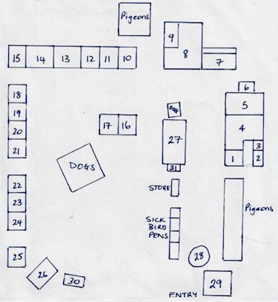
Table 1 includes the number of sick and clinically normal birds by cage number. Figure 1 includes a map of the poultry yard.
| Cage # | Description | Cage # | Description |
|---|---|---|---|
| 1 | 2 OK; 1 sick | 17 | 3 OK; 1 sick; 1 dead 9/5/23 |
| 2 | 1 OK | 18 | 2 OK; 1 sick |
| 3 | 1 sick - back from show on weekend | 19 | 6 OK; 1 sick |
| 4 | 8 OK; 1 sick; 2 dead | 20 | 3 OK |
| 5 | 3 OK; 2 sick (1 for PM); 4 died in cage in April | 21 | 3 OK |
| 6 | 1 OK | 22 | 4 OK |
| 7 | 6 OK (homebred); 15 bought in birds died Feb | 23 | Empty, pair died |
| 8 | 3 OK; 1 sick; 1 dead; 6 died recently | 24 | Empty, all died Feb except 1 hen now cage 28 |
| 9 | 6 OK; 3 sick (1 for PM) | 25 | 4 OK |
| 10 | 3 OK | 26 | 2 Guinea Fowl OK |
| 11 | 3 OK | 27 | 3 OK; 1 sick |
| 12 | 2 OK; 1 dead | 28 | 3 OK |
| 13 | Empty, 2 went to cage 5, 4 died here | 29 | 13 OK; 3 sick |
| 14 | Pigeons | 30 | 1 OK |
| 15 | 4 OK; 1 sick (for PM) | 31 | 1 OK |
| 16 | 4 OK |
CLINICAL HISTORY
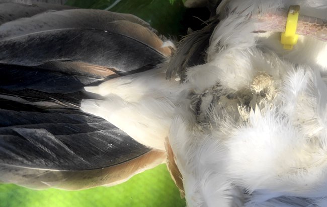
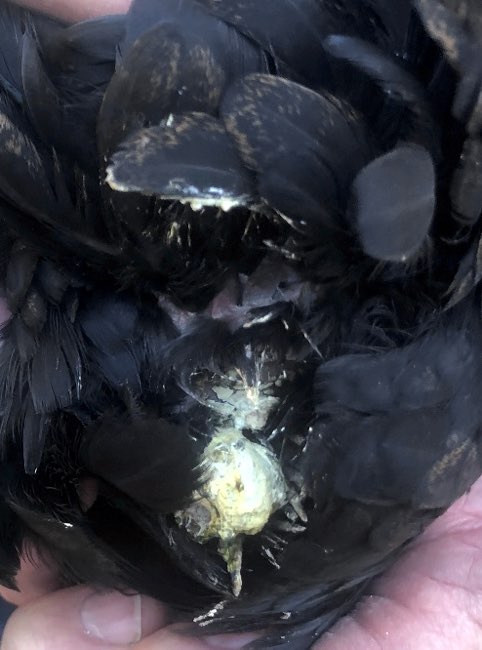
In the preceding months the owner had noticed numerous juvenile and adult birds becoming unwell and then dying. Commonly, he noted that the birds looked depressed, displayed ruffled plumage, lost condition and had eye discharge. Their combs would become dark in colour (purple). Some poultry also had white faeces accumulating around their vent (Figure 2). He noted that eye discharge was white and thick. Some birds appeared to be unwell for 1-5 days before death, yet others were found dead with no apparent signs of sickness.
He also reported deaths amongst the hatchlings. The chicks were hatched out and taken to rear, but all died within a week. The chicks became fluffed up and had watery ocular discharge prior to dying.
CLINICAL EXAMINATION
At the time of the property visit there were approximately 107 live poultry and, of these, at least 15 appeared unwell. There were an additional two dead birds. There were also numerous pigeons, but they were not counted nor examined as none were sick and there had been no unusual mortalities among these birds.
Four live birds were selected for clinical examination.
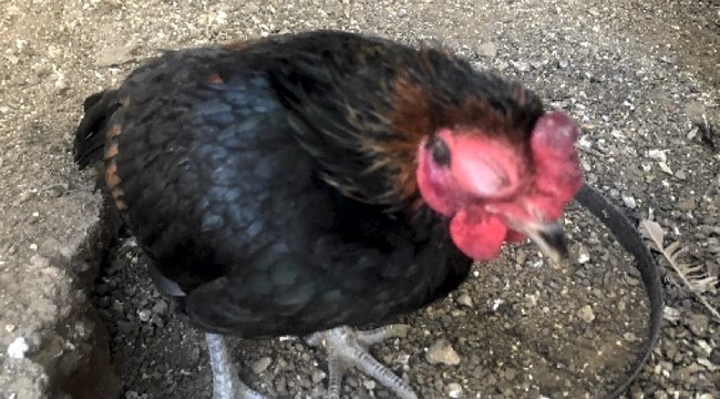
All birds were in good body condition but had darker-than-normal wattles and the wattles were not as erect as normal. Three birds had white material around their vents.
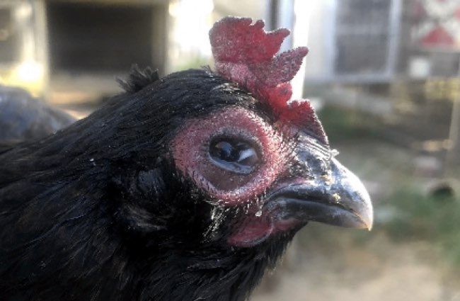
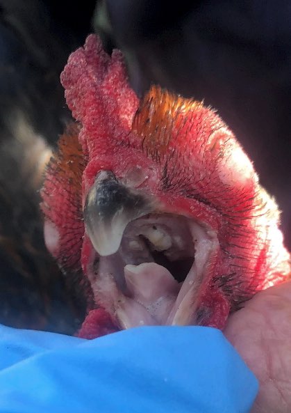
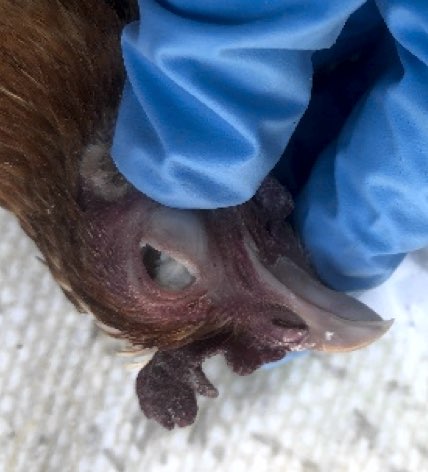
All birds had a white, caseous ocular discharge that almost appeared like a diphtheritic membrane across both eyes. They also had noticeable swelling of the tissues around both eyes (Figures 3 and 4).
One bird that was examined had a creamy coloured caseous material in the choanal slit (Figure 5).
No coughing or sneezing was heard when walking around the poultry yard.
POST-MORTEM EXAMINATION AND SAMPLE COLLECTION
Four birds were selected for post-mortem. One bird had died overnight (Bird 1), and one bird was euthanised just prior to post-mortem (Bird 2) to ensure fresh samples could be collected. Fresh and fixed samples were taken to be sent to Elizabeth Macarthur Agricultural Institute (EMAI). The other two birds (Birds 3 and 4) were euthanised and sent as whole birds so post-mortems could be conducted by pathologists at EMAI.
GROSS POST-MORTEM FINDINGS
Bird 1 - The only obvious findings on post-mortem was a white, caseous exudate found in the choanal slit, as well as white, caseous material across the cornea (Figure 6). There was no diphtheritic membrane or evidence of inflammation in the trachea. Grossly the bird appeared normal. Fresh and fixed samples were sent to EMAI.
Bird 2 - Again, the only obvious lesion seen on post-mortem was a white, caseous exudate in the choanal slit. This area was swabbed and the swab placed in bacterial swab media. A sample of the caseous exudate was placed in PBSG. Grossly, the bird appeared normal. Fresh and fixed samples were sent to EMAI.
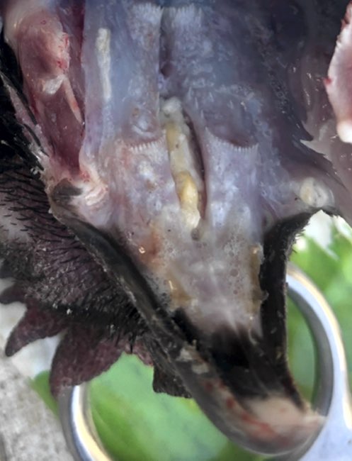
Birds 3 and 4 - The main gross post-mortem finding in both birds sent to EMAI was the presence of caseous material in the choanal slit (Figure 7). Also, Bird 4 had small, discrete white nodules (1-2mm) throughout the oesophagus.
DIFFERENTIAL DIAGNOSIS
- Avian influenza (AI)
- Infectious laryngotracheitis (ILT) (herpesvirus)
- Infectious coryza (Avibacterium paragallinarum)
- Chlamydia psittaci (psittacosis); Chlamydia gallinacean
- Fowl pox
- Mycoplasma gallisepticum
- Newcastle Disease
- Vitamin A deficiency
The first step was to rule out infectious causes as the owner had been to the local agricultural show with a bird in the preceding week. Avian influenza is a notifiable disease in NSW and needed to be ruled out as a cause of the morbidity and mortality.
LABORATORY TESTING
Samples were sent to EMAI.
The tests deemed appropriate to conduct by the pathologist were:
- Histopathology
- Bacteriology
- Chlamydia PCR
- Mycoplasma gallisepicum and Mycoplasma synoviae duplex qPCR
- Newcastle Disease Virus Real Time PCR
- ILT Real Time PCR
- Influenza A matrix PCR
LABORATORY RESULTS
The histopathology findings from the oesophagus revealed squamous gland metaplasia of the mucus glands, which is characteristic of vitamin A deficiency (Figure 8).
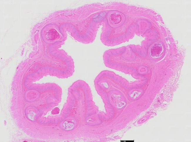
There was also evidence of mixed inflammation in the heart. Vitamin A deficiency does not cause this change directly but it does impair immune function. As such, the birds would be more susceptible to opportunistic infections, which could explain the inflammatory changes seen histopathologically in the heart.
The results for avian influenza A, Newcastle disease, ILT and Chlamydia psittaci were all negative.
Mycoplasma synoviae was detected by PCR. Infection with Mycoplasma spp. is common in backyard poultry flocks with clinical signs of infection commonly due to poor nutrition and husbandry. In this case any clinical disease due to the presence of M. synoviae would have occurred secondarily to the vitamin A deficiency.
Pseudomonas aeruginosa was also isolated from the sinus. Infection with this organism would have been secondary to the vitamin A deficiency and the associated immunocompromise.
DISCUSSION
Vitamin A deficiency in poultry is rarely seen in commercial operations. It is more common in backyard poultry operations as birds may be provided with home-mixed diets deficient in vitamin A and may not have access to areas for grazing. In this case the poultry owner had moved away from feeding commercially available poultry feed mixes to reduce feeding costs. In doing so he inadvertently caused hypovitaminosis A in his flock.
This case proved to be an interesting investigation as it is uncommon for many mixed animal veterinarians to investigate a poultry case. An understanding of vitamin A deficiency in poultry is important for mixed animal veterinarians as the keeping of poultry on a small scale in many towns and cities is becoming increasingly popular. Having knowledge of the differential diagnoses that a possible case of hypovitaminosis A raises, is important as many are notifiable diseases in Australia, as well as some having zoonotic potential.
REFERENCES
- Shastak Y & Pelletier W (2023) Nutritional Balance Matters: Assessing the Ramifications of Vitamin A Deficiency on Poultry Health and Productivity Poultry 2(4): 493-515 doi.org
- Khan RU, Khan A, Naz S, Ullah Q, Puvača N, Laudadio V, Mazzei D, Seidavi A, Ayasan T & Tufarelli V (2023) Pros and Cons of Dietary Vitamin A and Its Precursors in Poultry Health and Production: A Comprehensive Review Antioxidants (Basel) 12(5) : 1131
- Korver D (2024) Vitamin Deficiencies in Poultry MSD Manual Veterinary Manual www.msdvetmanual.com
- Davies M, Slattery S and Ellem J (2019) Vitamin A Deficiency in Sheep and Cattle in North-Western NSW Flock and Herd www.flockandherd.net.au
- Beveridge WIB and Hart L (1985) Viral, Bacterial and Fungal Diseases in Poultry Animal Health in Australia Vol 7