CASE NOTES
Lameness and foot lesions in Dorper sheep
Jillian Lawrence, University of Sydney Intern and Libby Read, District Veterinarian, North West LHPA
Posted Flock & Herd June 2011
Introduction
Three cases of lameness in Dorper sheep were investigated by the North West Livestock Health and Pest Authority (NWLHPA) during December 2010 and January 2011. Weather conditions in northwest NSW during December had delivered high levels of rainfall, resulting in flooding, followed by periods of high temperatures. This allowed lush pasture growth through December and January. Prolonged exposure to warm, humid conditions can predispose sheep to lameness (Hodgkinson 2010; Wassink et al. 2004). The differential diagnoses for lameness in sheep following wet, hot conditions include footrot, interdigital dermatitis, foot abscess and strawberry footrot (Hodgkinson 2010; Moriello 2008). NSW is currently declared a protected zone for footrot and virulent footrot has not been implicated in an outbreak of lameness in the NWLHPA for around 20 years (Slattery, S 2011, pers comm., 10th January). However, it is important to exclude this possibility during favourable environmental conditions.
The following three cases illustrate the similar presentations of lameness and appearance of foot lesions in Dorper sheep that can be attributed to varying causes. This presents a challenge for disease recognition by the producer and accurate diagnosis by the veterinarian. The number of cases, limited to Dorper breed sheep, over a short time period in the Narrabri region also raises the question of the possible predisposition to lameness of Dorper sheep following a wet event.
CASE 1
History
A NWLHPA District Veterinarian (DV) was called out to a small Dorper stud enterprise near Narrabri to examine a mob of severely lame Dorper sheep.
30 ewes had been introduced from Western Australia in October and November 2010. A single neighbouring property also ran sheep. However, there have been no known strays for a number of months. The affected mob had been grazing the same paddock for several months, comprising tall tropical perennial pasture with large areas of pooled water in recent weeks. The paddock contained Tribulus sp. which, combined with damp conditions, was suspected by the owner to have led to physical damage of the feet.
Lameness was noticed by the producer in one or two ewes, from a mob of 100, two weeks prior to the property visit. This number increased gradually to 50% of the mob identified to be lame during the property visit on 6 January 2011.
Clinical examination
Thirty acutely lame ewes were observed, and six ewes displaying the highest degree of lameness were examined.
All ewes were reluctant to walk or stand and some held individual feet off the ground. Of the ewes that were examined, all had Score 2 lesions in one to four feet. The interdigital skin was red and inflamed and a moist, pasty exudate was present at the skin-horn junction. The lesions had an anaerobic odour. The ewe suffering the most severe lameness had two feet with distinct lines of moisture and inflammation at the skin-horn junction, one of these feet had small maggots present. None of the sheep examined had swollen joints. There was no evidence of damage due to Tribulus sp.
Six swabs of the interdigital skin were taken and stored in footrot media for laboratory diagnosis.

Images 1 to 3: Score 2 lesions with moist exudate
Photos: Shaun Slattery, SDV NWLHPA
Treatment advice
Until laboratory testing determined whether virulent footrot was present, the producer was advised against selling any rams and to keep the mobs on the property separated, in their present paddocks. On 7 January 2011, another fly-struck sheep was discovered by the producer. The owner was advised to begin footbathing all affected animals with a 10% solution of zinc sulphate.
Footbathing was performed on 8 January 2011 and two days later it was reported that this had resolved almost all lameness in the mob.
Laboratory findings / diagnosis
Culture and gel diffusion test revealed benign Dichelobacter nodosus.
CASE 2
History
On 7 January 2011 a property visit was made to a Dorper stud located near Wee Waa to investigate sheep with lesions around the feet and mouth, and lumpy wool in one lamb. The farmer was concerned about footrot and the purpose of the visit was footrot exclusion. The lesions had been noticed when the sheep were yarded for management purposes in the morning.
The sheep were grazing paddocks containing thistles which may have had an abrasive effect on the feet and muzzle, potentially predisposing to scabby mouth.
Clinical examination
Five sheep were examined.
Ewe 1: Lame, with an interdigital lesion on one foot only, consistent with a penetrating injury.
Ewe 2: Lame, with a crusting lesion at the coronary band of one foot, approximately 3cm in diameter. Nodular crusting lesions were also present on the muzzle. These lesions were consistent with contagious orf.

Image 4: Bleeding lesion at coronary band (Ewe 2)

Image 5: Nodular crusting lesions around muzzle (Ewe 2)
Lamb 1: Crusting lesions located at the coronary band of two feet. Nodular, non-crusting lesion present on the face. These lesions were consistent with contagious orf.
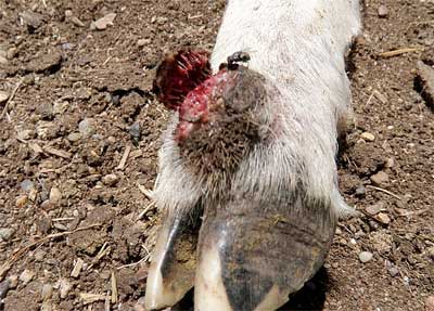
Image 6: Scab formation at coronary band (Lamb 1)
Lamb 2: Lesions on the head and dorsal area of the body consistent with severe dermatophilosis and contagious orf. The skin was severely inflamed, with areas of matted wool and scabs. These severe lesions were systemically debilitating.

Image 7: Severe dermatophilosis (Lamb 2)
Lamb 3: Severe contagious orf lesion on the head and face, the feet were unaffected.

Image 8: Severe contagious orf around muzzle
Photos: Libby Read, DV Narrabri/Walgett
Laboratory findings / diagnosis
Contagious orf (scabby mouth) was suspected due to the presence of characteristic lesions around both the muzzle and feet. This was complicated by a secondary bacterial infection (possibly dermatophilosis) in some sheep. Footrot was excluded due to the absence of interdigital lesions.
Treatment advice
The producer was advised that there is no effective treatment for scabby mouth; however any sheep that develop secondary infections should be treated with long acting penicillin at the recommended dose rate. Any sheep with lesions around the mouth or feet should not be sold until the condition resolves.
CASE 3
History
700 ewes were purchased from the Barraba region and trucked to a Dorper stud enterprise located near Narrabri in early December 2010. Upon arrival 70 animals were noticed to be lame. The lame sheep were immediately returned to the property from which they were purchased. During subsequent flock monitoring over the next few days, further animals were noticed to be lame. The proportion of lame ewes was approaching approximately 10% of the remaining flock. The flock were grazing on paddocks of native pasture 40cm high and 80% green.
A mob of 200 sheep were examined on 15 December 2010. A large proportion of lame sheep had joint swelling around the feet and purulent foot lesions. Some of these lesions had broken out to form draining tracts. Foot abscess was determined to be the most likely cause. Of the mob mustered for examination, four ewes were examined. Footrot was not suspected at this point due the absence of visible lesions between the toes. Severely lame animals were treated by the producer with long acting penicillin.
Six weeks later the producer was concerned that the foot abscess problem appeared unresolved. During the second property visit on 12 January 2011 the sheep appeared to be improving; the number of lame sheep had reduced to about 10% in the mob of 200 sheep.
Clinical examination
Several sheep appeared severely lame (non-weight bearing) in one limb, with swelling of the distal interphalangeal joint. These sheep had proliferative granulation tissue on the sole of the hoof, with a purulent exudate. These lesions were characteristic of foot abscess.

Image 9: Foot abscess
Four other ewes were examined. They were lame, but did not have visible swelling around the feet. The lesions were similar in each of these sheep. The interdigital skin was reddened and inflamed, with a moist exudate and some sloughing of the skin at the skin-horn junction. These lesions were consistent with footrot.


Images 10 and 11: Interdigital dermatitis with moist exudate
Photos: Ted Irwin, DV Warialda
Interdigital swabs were taken from the four ewes with these lesions and stored in footrot media for laboratory diagnosis.
Treatment advice
The farmer was advised to keep the affected mob separate to other sheep on the property until the lameness resolved or until lab results were available. There were no facilities for footbathing at the property.
Laboratory findings / diagnosis
Culture results and IntA PCR revealed benign Dichelobacter nodosus.
Discussion
Ovine footrot and interdigital dermatitis are considered to be related in their epidemiology. Interdigital dermatitis is caused by Fusobacterium necrophorum, an anaerobic organism, ubiquitous in the environment (Green & George 2008). Elevated moisture levels, high temperatures and dense pasture growth can create a favourable environment, allowing the organism to invade the feet of sheep (Graham & Egerton 1968). F. necrophorum has a necrotising effect on the interdigital skin, which causes interdigital dermatitis (Graham & Egerton 1968).
Dichelobacter nodosus is the causative agent of ovine footrot, an obligate anaerobic organism, which requires its host for survival (Roberts & Egerton 1969). It is thought that D. nodosus is able to colonize the feet of sheep subsequent to interdigital dermatitis infection with F. necrophorum. Virulent strains of D. nodosus cause underrunning of the horn of the hoof, resulting in lameness (Graham & Egerton 1968). D. nodosus may remain dormant in affected flocks and particular environmental conditions are required for the expression and spread of virulent footrot (Green & George 2008). The recent weather conditions experienced in the NWLHPA were suitable for virulent footrot to occur.
The aetiology of foot abscess in sheep is also linked to ovine interdigital dermatitis and F. necrophorum. Foot abscess occurs when F. necrophorum invades the interdigital skin, and extends below the basal layers into the distal interphalangeal joint to produce a suppurative arthritis (Corner, Collins & Vaughn 1996). Additional organisms are able to colonize the area and have been implicated in foot abscess, predominantly Corynebacterium pyogenes (Corner, Collins & Vaughn 1996; West 1983). In contrast to interdigital dermatitis and footrot, joint swelling is a feature of foot abscess, which was observed in Case 3 (Corner Collins & Vaughn 1996).
Contagious orf (scabby mouth) is a contagious viral disease of sheep with zoonotic potential. Sheep that are grazing abrasive feed such as stubble, T. terrestris or thistles are most susceptible and this is often when outbreaks occur (Stone 2007). Abrasive plants can cause damage to the mouth, feet, legs, teats and poll, allowing entry of the virus (Plant 2004). Lesions on the feet often result after animals have been grazing damp paddocks containing thistles (Plant 2004). This was the suspected initiating cause in Case 2. Contagious orf can be complicated by secondary bacterial infection in severe cases (Plant 2004), which occurred in one lamb in Case 2. Secondary infection with dermatophilosis can result in strawberry footrot (Moriello 2008).
Strawberry footrot (dermatophilosis) has a similar appearance to the lesions produced by contagious orf; however they are localized to the feet and coronet. The causative organism Dermatophilus congolensis produces an exudative dermatitis with scab formation (Moriello 2008). Often the scabs peel off to reveal a red, bleeding lesion. Risk factors for the occurrence of strawberry footrot include prolonged wetting by rain; high humidity; high temperatures and ectoparasites (Moriello 2008). Epidemics occur during seasons of high rainfall (Moriello 2008).
Dorper sheep are recognised for their good carcass quality and production characteristics in a typically low rainfall, grazing environment (Milne 2000). They are also known for being a hardy, low maintenance breed with greater resistance to some diseases than other sheep breeds (Schoeman 2000; Milne 2000). In northwest NSW, Dorpers are an increasingly popular meat breed as they are suitable for the environment and do not require frequent shearing. Dorpers originate in arid to semi-arid conditions and are not historically exposed to wet conditions (Milne 2000). Although there are claims that Dorpers are able to adapt to wet climates in South Africa (SA Dorper Breeder's Association 2011), there is minimal literature describing lameness in Dorpers subsequent to periods of high rainfall and temperatures.
The outbreaks of lameness observed in the NWLHPA suggest that Dorpers are susceptible to diseases affecting the foot including interdigital dermatitis, footrot, foot abscess and contagious orf; especially during prolonged warm and wet conditions. Lameness can cause significant production losses and compromise welfare. Appropriate flock management and monitoring during warm and wet environmental conditions could minimise the effects and incidence of lameness (Wassink et al. 2010).
The three cases presented here highlight the value of thorough feet examinations when sheep present for lameness or foot lesions following favourable environmental conditions for footrot, interdigital dermatitis, foot abscess, strawberry footrot and contagious orf. The lesions caused by these diseases can have a similar appearance (Kaler & Green 2008) and may be complicated by the presence of a concurrent disease process, as occurred in Cases 2 and 3. This can make it difficult to distinguish the cause of lameness or foot lesions. Sheep displaying a range of clinical signs, from non-lame to severely lame, with or without joint swelling, should be examined in order to make an accurate diagnosis in the field, or for appropriate samples to be taken (Winter 2004). In Case 3, some sheep showing signs of interdigital dermatitis may have been overlooked as only those sheep with joint swelling were examined. Several sheep showing a range of clinical signs were examined in Case 2. This allowed footrot to be confidently ruled out and scabby mouth to be considered more likely than strawberry footrot as the primary cause.
The early identification and diagnosis of ovine lameness is important in order to implement effective, economical treatment. In the case of virulent footrot in NSW, quarantine and an eradication plan is required to be implemented. This process would be labour intensive and costly to the producer. If recognised early, the extent of the outbreak would be minimised thus reducing not only time and financial costs but production losses as well. Similar principles apply to the early implementation of a treatment plan for foot abscess or interdigital dermatitis. Although there is no effective treatment for scabby mouth (Stone 2007), correct and timely diagnosis can limit the spread of disease both on farm and between properties. The ability to protect the producer's reputation is also an important factor, which was considered in each of these cases, as they occurred on stud properties.
It can be concluded from these cases that Dorper sheep are susceptible to lameness subsequent to prolonged exposure to warm, damp conditions. This raises management issues for Dorper sheep during periods of wet weather extremes in a semi-arid environment. The early recognition of lameness by the producer and accurate diagnosis by the veterinarian will provide the best opportunity to manage and treat the causative problem.
References
- Corner LA, Collins ND & Vaughan JA (1996) An experimental ovine foot abscess model using a Fusobacterium necrophorum biotype AB Veterinary Microbiology 48(1-2):1-7
- Graham NP & Egerton JR (1968) Pathogenesis of ovine foot-rot: the role of some environmental factors, Australian Veterinary Journal 44(5):235-240
- Green Le & George TRN (2008) Assessment of current knowledge of footrot in sheep with particular reference to Dichelobacter nodosus and implications for elimination or control strategies for sheep in Great Britain The Veterinary Journal 175(2):173-180
- Hodgkinson O (2010) The importance of feet examination in sheep health management Small Ruminant Research 92(1-3):67-71
- Kaler J & Green LE (2008) Naming and recognition of six foot lesions of sheep using written and pictorial information: A study of 809 English sheep farmers, Preventive Veterinary Medicine 83(1):52-64
- Milne C (2000) The history of the Dorper sheep Small Ruminant Researc, 36(2):99-102
- Moriello K (2008) Dermatophilosis: Introduction, Merck Veterinary Manual, viewed 8th January 2011 www.merckvetmanual.com
- Plant JW (2004) Sheep health: scabby mouth, Agfact, NSW Department of Agriculture
- Roberts DS & Egerton JR (1969) The aetiology and pathogenesis of ovine foot-rot. II. The pathogenic association of Fusiformis nodosus and F. Necrophorus Journal of Comparative Pathology 79>(2):217-227
- Schoeman SJ (2000) A comparative assessment of Dorper sheep in different production environments and systems Small Ruminant Research 36(2):137-146
- South African (SA) Dorper Breeder's Association (2011) Breed Characteristics, viewed 17th January 2011 www.dorpersa.co.za
- Stone N (2007) Scabby Mouth (Orf) A Disease of Sheep and Goats, Agriculture notes, Department of Primary Industries, Victoria
- Wassink GJ, George TRN, Kaler J & Green LE (2010) Footrot and interdigital dermatitis in sheep: Farmer satisfaction with current management, their ideal management and sources used to adopt new strategies Preventive Veterinary Medicine 96(1-2):65-73
- Wassink GJ, Grogono-Thomas R, Moore LJ & Green LE (2004) Risk factors associated with the prevalence of interdigital dermatitis in sheep from 1999 to 2000 The Veterinary Record 154(18):551-555
- West D (1983) A study of naturally occurring cases of ovine foot abscess in New Zealand New Zealand Veterinary Journal 31(9):152-156
- Winter A (2004) Lameness in Sheep 1. Diagnosis In Practice 26(2):58-63