CASE NOTES
UROLITHIASIS DUE TO AN UNBALANCED DIET IN A SPANISH LAMB FEEDLOT
Carlos Alcay, Luis Miguel Ferrer, Juan Jose Ramos, Marta Ruiz de Arcaute, Marta Borobia and Delia Lacasta, Animal Pathology Department, Veterinary Faculty, University of Zaragoza, Spain
Posted Flock & Herd March 2012
History
In recent months (September-November, 2011) there have been sporadic deaths in Rasa Aragonesa feedlot lambs on farms near Zaragoza in Spain. As a similar outbreak in 2010 caused a morbidity of 2% and mortality of 1%, two affected lambs were presented to the Small Ruminant External Consultancy Practice of the Veterinary Faculty of Zaragoza (Spain) to be evaluated. All these lambs were fed with commercial pellets and the factory told to the farmer that no changes had been made in the components of the ration. However, after this talk no more clinical cases appeared.
Clinical examination
The lambs had anorexia, symptoms indicative of colic pain, dysuria and extensive oedema of the lower abdomen and particularly the prepuce (Figure 1). A summary of clinical parameters is presented (Table 1).

Figure 1: swelling of the prepuce in a feedlot lamb
| Lamb 1 | Lamb 2 | |
|---|---|---|
| Body condition scoring | 3 | 3 |
| Temperature | 38.1 °C | 38.5 °C |
| Heart rate | 128 bpm | 104 bpm |
| Respiratory rate | 34 rpm | 44 rpm |
| Symptoms | Ventral and testicular edema | Inflammation and pain in preputial area |
Table 1: clinical parameters in two lambs with swelling of the prepuce
Laboratory tests & findings
Tests included haematology (complete blood count), biochemical analysis and a smear of the prepuce.
Ancillary findings were that Lamb 1 had a slightly elevation of haemoglobin level and the haematocrit, with no abnormalities in blood from Lamb 2. Biochemistry identified that Lamb 1 had elevated total protein and very high urea and creatinine levels, with elevations in urea and creatinine levels in Lamb 2. No abnormalities of the smear from the prepuce were present in Lamb 1 although struvite crystals were found in the smear from Lamb 2.
Post-mortem findings
Lamb 1 deteriorated with anuria, anorexia, stupor and widespread ventral edema was necropsied. Pathological findings included subcutaneous edema of the prepuce, glomerulonephritis with mineral precipitation, severe locally extensive haemorrhage of the urethra, plus a large calculus and some small calculi blocking the urethra (Figures 2 and 3), identified as struvite crystals in a urine sample.

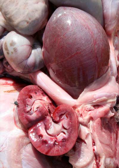
Figures 2 & 3: haemorrhage of the urethra, distension of the urinary bladder and renal pelvis from Lamb 1


Figures 4 & 5: urine was obtained from urinary bladder exmained microscopically revealed the presence of struvite crystals from Lamb 1
Diagnosis
Consideration of the history, clinical examination and the laboratory and necropsy findings indicated a diagnosis of urolithiasis.
An imbalance in levels of Ca & P in the diet predisposes to struvite crystal precipitation. Struvite calculi form when crystals join and accumulate in the urinary bladder, blocking the urethra, with subsequent rupture. Although urolithiasis can be present in females, symptoms mainly develop in males due to the anatomy of the urethra.
Therapy
Lamb 2 was treated with intravenous antispasmodics and analgesics (dipirona + hioscina) every 6-12 hours. Ventral edema disappeared progressively during the following days. Total recovery occurred within a week.
When a calculus completely obstructs the urethra, it may be removed by surgical amputation of vermiform plexus, where calculi are often located. However urethrectomy and exteriorization of the urethra is costly and usually impractical for sheep production.
Preventative recommendations
Recommendations were to address the dietary ration and provide balanced levels of calcium and phosphorus (at a 2:1 ratio), plus guarantee water availability and acidify the urine pH with the addition of ammonium chloride (0.4%) in the diet.